Brain Networks Involved in Sensory Perception in Parkinson's Disease: A Scoping Review
- PMID: 38002513
- PMCID: PMC10669548
- DOI: 10.3390/brainsci13111552
Brain Networks Involved in Sensory Perception in Parkinson's Disease: A Scoping Review
Abstract
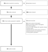
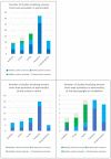
Parkinson's Disease (PD) has historically been considered a disorder of motor dysfunction. However, a growing number of studies have demonstrated sensory abnormalities in PD across the modalities of proprioceptive, tactile, visual, auditory and temporal perception. A better understanding of these may inform future drug and neuromodulation therapy. We analysed these studies using a scoping review. In total, 101 studies comprising 2853 human participants (88 studies) and 125 animals (13 studies), published between 1982 and 2022, were included. These highlighted the importance of the basal ganglia in sensory perception across all modalities, with an additional role for the integration of multiple simultaneous sensation types. Numerous studies concluded that sensory abnormalities in PD result from increased noise in the basal ganglia and increased neuronal receptive field size. There is evidence that sensory changes in PD and impaired sensorimotor integration may contribute to motor abnormalities.
Keywords: Parkinson’s; basal ganglia; brain network; functional magnetic imaging; microelectrode; positron emission tomography; sensorimotor; sensory.
Conflict of interest statement
The authors declare no conflict of interest.
Figures


Similar articles
-
Fast Intracortical Sensory-Motor Integration: A Window Into the Pathophysiology of Parkinson's Disease.Front Hum Neurosci. 2019 Apr 8;13:111. doi: 10.3389/fnhum.2019.00111. eCollection 2019. Front Hum Neurosci. 2019. PMID: 31024277 Free PMC article.
-
Sensorimotor integration in movement disorders.Mov Disord. 2003 Mar;18(3):231-240. doi: 10.1002/mds.10327. Mov Disord. 2003. PMID: 12621626 Review.
-
Functional brain imaging: an evidence-based analysis.Ont Health Technol Assess Ser. 2006;6(22):1-79. Epub 2006 Dec 1. Ont Health Technol Assess Ser. 2006. PMID: 23074493 Free PMC article.
-
Abnormal phasic activity in saliency network, motor areas, and basal ganglia in Parkinson's disease during rhythm perception.Hum Brain Mapp. 2019 Feb 15;40(3):916-927. doi: 10.1002/hbm.24421. Epub 2018 Oct 29. Hum Brain Mapp. 2019. PMID: 30375107 Free PMC article.
-
Proprioceptive dysfunction in focal dystonia: from experimental evidence to rehabilitation strategies.Front Hum Neurosci. 2014 Dec 9;8:1000. doi: 10.3389/fnhum.2014.01000. eCollection 2014. Front Hum Neurosci. 2014. PMID: 25538612 Free PMC article. Review.
Cited by
-
A distinct circuit for biasing visual perceptual decisions and modulating superior colliculus activity through the mouse posterior striatum.bioRxiv [Preprint]. 2024 Jul 31:2024.07.31.605853. doi: 10.1101/2024.07.31.605853. bioRxiv. 2024. PMID: 39372791 Free PMC article. Preprint.
-
Hypo-connectivity of the primary somatosensory cortex in Parkinson's disease: a resting-state functional MRI study.Front Neurol. 2024 Apr 30;15:1361063. doi: 10.3389/fneur.2024.1361063. eCollection 2024. Front Neurol. 2024. PMID: 38746656 Free PMC article.
-
Feasibility of a Community-Based Boxing Program with Tailored Balance Training in Parkinson's Disease: A Preliminary Study.Brain Sci. 2025 Aug 13;15(8):858. doi: 10.3390/brainsci15080858. Brain Sci. 2025. PMID: 40867189 Free PMC article.
-
Redefining Non-Motor Symptoms in Parkinson's Disease.J Pers Med. 2025 Apr 26;15(5):172. doi: 10.3390/jpm15050172. J Pers Med. 2025. PMID: 40423044 Free PMC article. Review.
-
Distinct patterns of visuo-tactile and visuo-motor body-related integration in Parkinson's disease.Sci Rep. 2025 Jul 31;15(1):27923. doi: 10.1038/s41598-025-08965-5. Sci Rep. 2025. PMID: 40744963 Free PMC article.
References
-
- Parkinson J. An Essay on the Shaking Palsy. Whittingham and Rowland for Sherwood, Neely and Jones; London, UK: 1817.
-
- NCCC (National Collaborating Centre for Chronic Conditions) (UK) Parkinson’s Disease: National Clinical Guideline for Diagnosis and Management in Primary and Secondary Care. Royal College of Physicians; London, UK: 2006. - PubMed
Publication types
LinkOut - more resources
Full Text Sources

