

Neural Mechanisms of Neuro-Rehabilitation Using Transcranial Direct Current Stimulation (tDCS) over the Front-Polar Area
- PMID: 38002563
- PMCID: PMC10670271
- DOI: 10.3390/brainsci13111604
Neural Mechanisms of Neuro-Rehabilitation Using Transcranial Direct Current Stimulation (tDCS) over the Front-Polar Area
Abstract
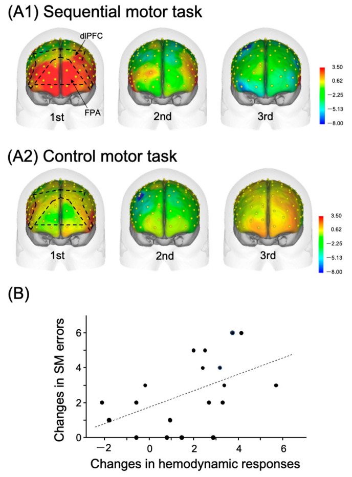
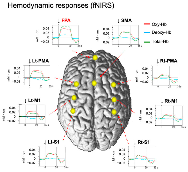
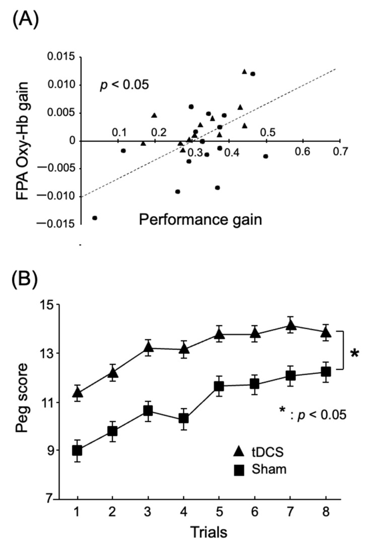
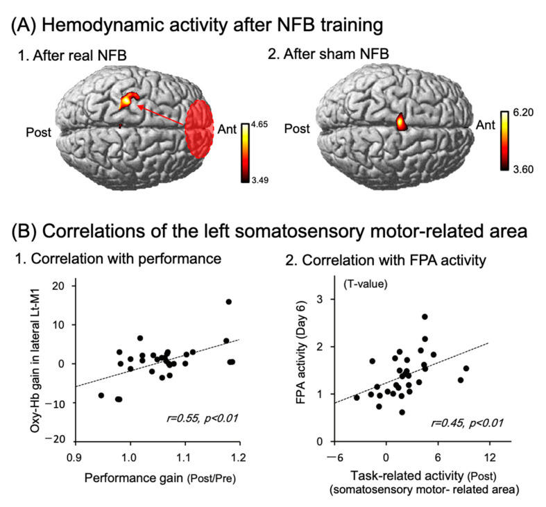
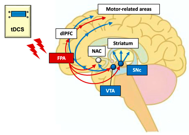
Transcranial direct current stimulation (tDCS) is a noninvasive brain stimulation (NIBS) technique that applies a weak current to the scalp to modulate neuronal excitability by stimulating the cerebral cortex. The technique can produce either somatic depolarization (anodal stimulation) or somatic hyperpolarization (cathodal stimulation), based on the polarity of the current used by noninvasively stimulating the cerebral cortex with a weak current from the scalp, making it a NIBS technique that can modulate neuronal excitability. Thus, tDCS has emerged as a hopeful clinical neuro-rehabilitation treatment strategy. This method has a broad range of potential uses in rehabilitation medicine for neurodegenerative diseases, including Parkinson's disease (PD). The present paper reviews the efficacy of tDCS over the front-polar area (FPA) in healthy subjects, as well as patients with PD, where tDCS is mainly applied to the primary motor cortex (M1 area). Multiple evidence lines indicate that the FPA plays a part in motor learning. Furthermore, recent studies have reported that tDCS applied over the FPA can improve motor functions in both healthy adults and PD patients. We argue that the application of tDCS to the FPA promotes motor skill learning through its effects on the M1 area and midbrain dopamine neurons. Additionally, we will review other unique outcomes of tDCS over the FPA, such as effects on persistence and motivation, and discuss their underlying neural mechanisms. These findings support the claim that the FPA could emerge as a new key brain region for tDCS in neuro-rehabilitation.
Keywords: Parkinson’s disease; neural mechanisms; the frontal pole area; transcranial direct current stimulation.
Conflict of interest statement
The authors declare no conflict of interest.
Figures
Similar articles
-
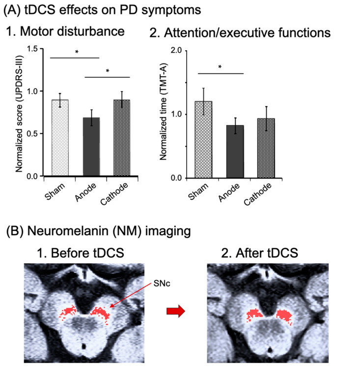
Effects of Transcranial Direct Current Stimulation (tDCS) Over the Frontal Polar Area on Motor and Executive Functions in Parkinson's Disease; A Pilot Study.Front Aging Neurosci. 2018 Jul 30;10:231. doi: 10.3389/fnagi.2018.00231. eCollection 2018. Front Aging Neurosci. 2018. PMID: 30104971 Free PMC article.
-
A Parkinson's disease patient displaying increased neuromelanin-sensitive areas in the substantia nigra after rehabilitation with tDCS: a case report.Neurocase. 2021 Oct;27(5):407-414. doi: 10.1080/13554794.2021.1975768. Epub 2021 Sep 10. Neurocase. 2021. PMID: 34503372
-
The Effect of a Single Session of Non-Invasive Brain Stimulation on Balance in Healthy Individuals: A Systematic Review and Best Evidence Synthesis.Brain Connect. 2021 Nov;11(9):695-716. doi: 10.1089/brain.2020.0872. Epub 2021 Sep 3. Brain Connect. 2021. PMID: 33798002
-
Transcranial direct-current stimulation combined with attention increases cortical excitability and improves motor learning in healthy volunteers.J Neuroeng Rehabil. 2020 Feb 19;17(1):23. doi: 10.1186/s12984-020-00665-7. J Neuroeng Rehabil. 2020. PMID: 32075667 Free PMC article.
-
Frontal Transcranial Direct Current Stimulation as a Potential Treatment of Parkinson's Disease-Related Fatigue.Brain Sci. 2021 Apr 8;11(4):467. doi: 10.3390/brainsci11040467. Brain Sci. 2021. PMID: 33917684 Free PMC article. Review.
Cited by
-
Cognitive and neurophysiological effects of bilateral tDCS neuromodulation in patients with minimally conscious state.Sci Rep. 2025 Apr 24;15(1):14389. doi: 10.1038/s41598-025-99591-8. Sci Rep. 2025. PMID: 40274956 Free PMC article. Clinical Trial.
References
-
- Antal A., Alekseichuk I., Bikson M., Brockmöller J., Brunoni A., Chen R., Cohen L., Dowthwaite G., Ellrich J., Flöel A., et al. Low intensity transcranial electric stimulation: Safety, ethical, legal regulatory and application guidelines. Clin. Neurophysiol. 2017;128:1774–1809. doi: 10.1016/j.clinph.2017.06.001. - DOI - PMC - PubMed
-
- Khedr E.M., Shawky O.A., El-Hammady D.H., Rothwell J.C., Darwish E.S., Mostafa O.M., Tohamy A.M. Effect of Anodal versus Cathodal Transcranial Direct Current Stimulation on Stroke Rehabilitation: A Pilot Randomized Controlled Trial. Neurorehabilit. Neural Repair. 2013;27:592–601. doi: 10.1177/1545968313484808. - DOI - PubMed
Publication types
Grants and funding
LinkOut - more resources
Full Text Sources
