

A Systematic Review and Illustrative Case Presentation of Low-Grade Myofibroblastic Sarcoma (LGMS) of the Extremities
- PMID: 38002641
- PMCID: PMC10672639
- DOI: 10.3390/jcm12227027
A Systematic Review and Illustrative Case Presentation of Low-Grade Myofibroblastic Sarcoma (LGMS) of the Extremities
Abstract
Introduction: Low-grade myofibroblastic sarcoma (LGMS) is a rare tumor entity which occurs in the subcutaneous and deep soft tissues; it is less common in the bone with a predilection for the extremities and the head and neck region. As confirming the diagnosis is difficult and treatment strategies are not standardized, we aimed to identify patient and tumor characteristics, and to summarize treatment strategies and their clinical outcomes to guide surgeons.
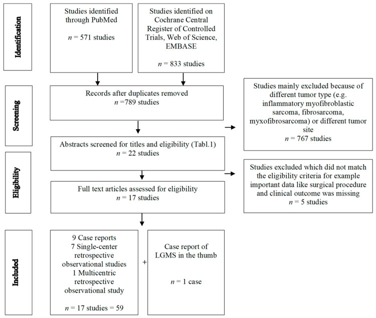
Methods: Included were full articles reporting patients with histology of LGMS in the extremities, excluding tumors of the trunk. All patients underwent surgery but with different extend, from marginal to wide resection. Included studies should inform about local recurrence, metastasis, or evidence of disease, depending on the surgical treatment. We conducted a structured search using MEDLINE (via PubMed), Web of Science, EMBASE and Cochrane Central Register of Controlled Trials (CENTRAL) to identify studies on low-grade myofibroblastic sarcoma of the extremities. Study designs like randomized controlled trials, systematic reviews, prospective trials, retrospective studies, and case reports were included. Prospective studies and comparative studies were not available at all. Therefore, meta-analysis was not possible and statistical analysis was purely descriptive.
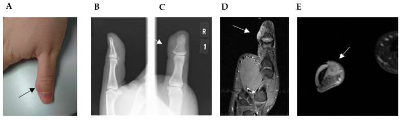
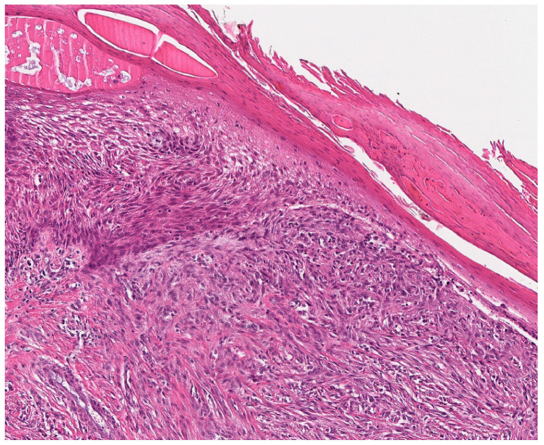
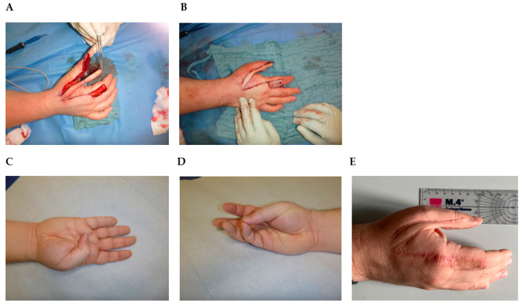
Results: Of the 789 studies identified from our initial search, 17 studies including 59 cases reported LGMS of the extremities with the surgical treatment and clinical outcome and were therefore analyzed. In addition, we present the rare case and surgical management of a 28-year-old male patient with residual LGMS of the thumb after an initial incomplete resection. The current literature suggests that a wide excision with R0 margins should be considered the standard treatment for LGMS. In cases where surgery leads to significant functional impairment, individual options like free tissue transfer from a donor site have to be considered. Therefore, we also present an illustrative case. For all selected case series and case reports, a high risk of confounding, selection bias, information bias, and reporting bias must be anticipated. Nevertheless, this systematic review provides a comprehensive overview on surgical treatment and clinical outcomes in LGMS surgery of the extremities.
Keywords: Holevich’s flap; LGMS; myofibrosarcoma; soft tissue sarcoma; thumb reconstruction surgery.
Conflict of interest statement
The authors declare no conflict of interest. The authors alone are responsible for the content and writing of the paper.
Figures
Similar articles
-
Low-grade myofibrosarcoma of the maxillary sinus: Two case reports.World J Clin Oncol. 2024 Apr 24;15(4):566-575. doi: 10.5306/wjco.v15.i4.566. World J Clin Oncol. 2024. PMID: 38689628 Free PMC article.
-
Low-grade myofibroblastic sarcoma of the levator scapulae muscle: a case report and literature review.BMC Musculoskelet Disord. 2020 Dec 10;21(1):836. doi: 10.1186/s12891-020-03857-3. BMC Musculoskelet Disord. 2020. PMID: 33302922 Free PMC article. Review.
-
Surgical treatment and long-term outcomes of low-grade myofibroblastic sarcoma: a single-center case series of 15 patients.World J Surg Oncol. 2021 Dec 7;19(1):339. doi: 10.1186/s12957-021-02454-5. World J Surg Oncol. 2021. PMID: 34872570 Free PMC article.
-
Indolent growth of low-grade myofibroblastic sarcoma of the cheek mimics benign lesions: A case report and literature review.Oncol Lett. 2017 Jun;13(6):4307-4314. doi: 10.3892/ol.2017.6020. Epub 2017 Apr 10. Oncol Lett. 2017. PMID: 28588708 Free PMC article.
-
Low-Grade Myofibroblastic Sarcoma of the Oral and Maxillofacial Region: An International Clinicopathologic Study of 13 Cases and Literature Review.Head Neck Pathol. 2023 Sep;17(3):832-850. doi: 10.1007/s12105-023-01577-3. Epub 2023 Aug 4. Head Neck Pathol. 2023. PMID: 37540486 Free PMC article. Review.
Cited by
-
Huge intermediate-grade myofibroblastic sarcoma in the retroperitoneum revealed by 18F-FDG PET/CT: a case report.Front Med (Lausanne). 2024 Sep 12;11:1461749. doi: 10.3389/fmed.2024.1461749. eCollection 2024. Front Med (Lausanne). 2024. PMID: 39328318 Free PMC article.
-
Low-Grade Myofibroblastic Sarcoma of the Acetabulum - A Rare Case with Review of Literature.J Orthop Case Rep. 2025 Jan;15(1):61-66. doi: 10.13107/jocr.2025.v15.i01.5126. J Orthop Case Rep. 2025. PMID: 39801859 Free PMC article.
-
Case Report: First case of low-grade myofibroblastic sarcoma of the vulva during pregnancy.Front Oncol. 2025 Jun 10;15:1577068. doi: 10.3389/fonc.2025.1577068. eCollection 2025. Front Oncol. 2025. PMID: 40556675 Free PMC article.
References
-
- Vasudev K.S., Harris M. A sarcoma of myofibroblasts: An ultrastructural study. Arch. Pathol. Lab. Med. 1978;102:185–188. - PubMed
-
- WHO Classification of Tumours Editorial Board . WHO Classification of Tumours: Soft Tissue and Bone Tumours. International Agency for Research on Cancer, IARD Press; Lyon, France: 2020.
Publication types
LinkOut - more resources
Full Text Sources
Research Materials
