

Case Report: Fetoscopic Laparoschisis (FETO-LAP)-A New Therapeutic Route to Explore for Fetuses with Severe Diaphragmatic Hernias
- PMID: 38002849
- PMCID: PMC10670710
- DOI: 10.3390/children10111758
Case Report: Fetoscopic Laparoschisis (FETO-LAP)-A New Therapeutic Route to Explore for Fetuses with Severe Diaphragmatic Hernias
Abstract
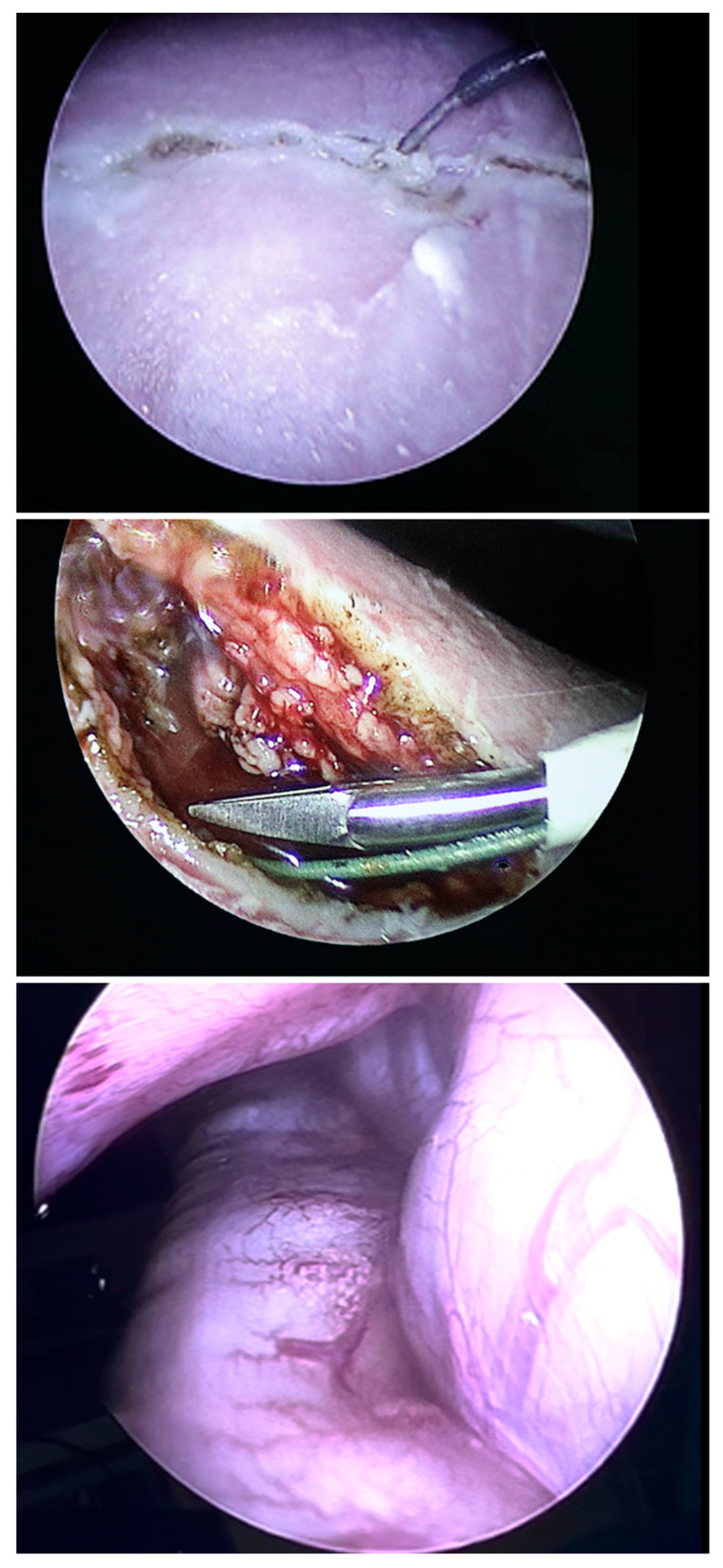
Background: The purpose of this report is to describe the seminal case of a near-term human fetus with a life-threatening left diaphragmatic hernia that underwent fetoscopic tracheal occlusion (FETO) combined with fetoscopic partial removal of herniated bowel from the fetal chest by fetoscopic laparoschisis (FETO-LAP).
Case summary: A life-threatening left diaphragmatic hernia (liver-up; o/e LHR of ≤25%; MRI lung volume ≤ 20%) was observed in a human fetus at 34 weeks of gestation. After counselling the mother about the high risks of postnatal demise if left untreated, the expected limitations of fetoscopic tracheal occlusion (FETO), and the previously untested option of combining FETO with fetoscopic laparoschisis, i.e., partial removal of the herniated bowel from the fetal chest (FETO-LAP), she consented to the latter novel treatment approach. FETO-LAP was performed at 36 + 5 weeks of gestation under general maternofetal anesthesia. Mother and fetus tolerated the procedure well. The neonate was delivered and the balloon removed on placental support at 37 + 2 weeks of gestation. On ECMO, a rapid increase in tidal volume was seen over the next eight days. Unfortunately, after this period, blood clots obstructed the ECMO circuit and the neonate passed away.
Discussion: This seminal case shows that in a fetus with severe left diaphragmatic hernia, partial removal of the herniated organs from the fetal chest is not only possible by minimally invasive fetoscopic techniques but also well tolerated. As the effect of FETO alone is limited in saving severely affected fetuses, combining FETO with fetoscopic laparoschisis (FETO-LAP) offers a new therapeutic route with multiple, potentially life-saving implications.
Keywords: FETO; diaphragmatic hernia; experimental surgery; fetal intervention; fetal surgery; fetoscopy; laparoschisis; new techniques; tracheal balloon occlusion.
Conflict of interest statement
The authors declare no conflict of interest.
Figures


Similar articles
-
[Fetoscopic tracheal occlusion for the treatment of severe congenital diaphragmatic hernia: preliminary results].Rev Esp Anestesiol Reanim. 2008 Aug-Sep;55(7):407-13. doi: 10.1016/s0034-9356(08)70611-x. Rev Esp Anestesiol Reanim. 2008. PMID: 18853678 Spanish.
-
Fetoscopic tracheal occlusion (FETO) for severe congenital diaphragmatic hernia: evolution of a technique and preliminary results.Ultrasound Obstet Gynecol. 2004 Aug;24(2):121-6. doi: 10.1002/uog.1711. Ultrasound Obstet Gynecol. 2004. PMID: 15287047
-
Sonographic pulmonary response after tracheal occlusion in fetuses with severe isolated congenital diaphragmatic hernia.J Clin Ultrasound. 2022 Feb;50(2):185-190. doi: 10.1002/jcu.23121. Epub 2022 Jan 12. J Clin Ultrasound. 2022. PMID: 35019149
-
Fetal intervention for congenital diaphragmatic hernia: the European experience.Semin Perinatol. 2005 Apr;29(2):94-103. doi: 10.1053/j.semperi.2005.04.006. Semin Perinatol. 2005. PMID: 16050527 Review.
-
Postnatal care setting and survival after fetoscopic tracheal occlusion for severe congenital diaphragmatic hernia: A systematic review and meta-analysis.J Pediatr Surg. 2022 Dec;57(12):819-825. doi: 10.1016/j.jpedsurg.2022.05.011. Epub 2022 May 19. J Pediatr Surg. 2022. PMID: 35680463
Cited by
-
Fetoscopic Endoluminal Tracheal Occlusion-Synergic Therapies in the Prenatal Treatment of Congenital Diaphragmatic Hernia.Int J Mol Sci. 2025 Feb 14;26(4):1639. doi: 10.3390/ijms26041639. Int J Mol Sci. 2025. PMID: 40004103 Free PMC article. Review.
References
-
- Ruano R., Yoshisaki C.T., Da Silva M.M., Ceccon M.E.J., Grasi M.S., Tannuri U., Zugaib M. A randomized controlled trial of fetal endoscopic tracheal occlusion versus postnatal management of severe isolated congenital diaphragmatic hernia. Ultrasound Obstet. Gynecol. 2012;39:20–27. doi: 10.1002/uog.10142. - DOI - PubMed
-
- Lynn D., Montgomery L.D., Belfort M.A., Saade G.R., Baker W., Pokorny W., Minifee P., Langston C., Jevon G., Van den Veyver I., et al. Iatrogenic gastroschisis decreases pulmonary hypoplasia in an ovine congenital diaphragmatic hernia modell. Fetal Diagn. Ther. 1995;10:119–126. doi: 10.1159/000264217. - DOI - PubMed
Publication types
LinkOut - more resources
Full Text Sources
Research Materials
Miscellaneous
