. 2023 Nov 9;24(22):16129.
doi: 10.3390/ijms242216129.

Evaluating Manganese, Zinc, and Copper Metal Toxicity on SH-SY5Y Cells in Establishing an Idiopathic Parkinson's Disease Model

Affiliations

Evaluating Manganese, Zinc, and Copper Metal Toxicity on SH-SY5Y Cells in Establishing an Idiopathic Parkinson's Disease Model

Sahar H Pradhan et al. Int J Mol Sci. .

Abstract

Parkinson's disease (PD) is a neurodegenerative condition marked by loss of motor coordination and cognitive impairment. According to global estimates, the worldwide prevalence of PD will likely exceed 12 million cases by 2040. PD is primarily associated with genetic factors, while clinically, cases are attributed to idiopathic factors such as environmental or occupational exposure. The heavy metals linked to PD and other neurodegenerative disorders include copper, manganese, and zinc. Chronic exposure to metals induces elevated oxidative stress and disrupts homeostasis, resulting in neuronal death. These metals are suggested to induce idiopathic PD in the literature. This study measures the effects of lethal concentration at 10% cell death (LC10) and lethal concentration at 50% cell death (LC50) concentrations of copper, manganese, and zinc chlorides on SH-SY5Y cells via markers for dopamine, reactive oxygen species (ROS) generation, DNA damage, and mitochondrial dysfunction after a 24 h exposure. These measurements were compared to a known neurotoxin to induce PD, 100 µM 6-hydroxydopamine (6-ODHA). Between the three metal chlorides, zinc was statistically different in all parameters from all other treatments and induced significant dopaminergic loss, DNA damage, and mitochondrial dysfunction. The LC50 of manganese and copper had the most similar response to 6-ODHA in all parameters, while LC10 of manganese and copper responded most like untreated cells. This study suggests that these metal chlorides respond differently from 6-ODHA and each other, suggesting that idiopathic PD utilizes a different mechanism from the classic PD model.

Keywords: SH-SY5Y; dose–response curves; hydroxydopamine; neurodegeneration; occupational exposure.


Conflict of interest statement

The authors declare no conflict of interest.

Figures

Figure 1
Figure 1
Dose–response curves for three chloride metals, (A) manganese chloride, (B) zinc chloride, and (C) copper chloride on SH-SY5Y cells for a 24 h exposure. Viability was determined via normalization against untreated cells. LC10 was determined to be a “sublethal concentration” (dotted), and LC50 was determined to be a “lethal concentration” (solid). The molarity equivalent of concentration is displayed in the left corner of each respective graph.
Figure 2
Figure 2
Relative H2O2 production was measured after a 24 h exposure to MnCl2, ZnCl2, and CuCl2 and 100 µM 6-ODHA. As a positive control, 50 µM menadione was used, and 1% PBS was used as a negative control. After 24 h, copper at lethal and sublethal concentrations had the highest effect, then zinc, and lastly, manganese at only the lethal concentration resulted in elevated ROS compared to the untreated (UT). Statistical significance codes: treatment vs. untreated control (UT, *) and positive control (6-ODHA, +). Asterisks or plus signs indicate p-value ranges: + 0.01–0.05, ++ 0.001–0.01, *** 0.0001 to 0.001, ****,++++ <0.0001.
Figure 3
Figure 3
Conserved dopamine neurotrophic factor (CDNF) concentration (pg/mL) after 24 h exposure to MnCl2, ZnCl2, and CuCl2 and 100 µM 6-ODHA. As a positive control, 50 µM menadione was used, and 1% PBS was used as a negative control. After 24 h exposure, ZnCl2 at lethal and sublethal concentrations had the highest effect, then CuCl2 and MnCl2. Statistical significance codes: treatment vs. untreated control (UT, *) and positive control (6-ODHA, +). Asterisks or plus signs indicate p-value ranges: *,+ 0.01–0.05, ++ 0.001–0.01, *** 0.0001 to 0.001, ****,++++ <0.0001.
Figure 4
Figure 4
8-hydroxy-2’-deoxyguanosine (8-OHdG) species were measured in digested DNA samples (pg/mL) after exposure to MnCl2, ZnCl2, and CuCl2 and 100 µM 6-ODHA. Use of 1% PBS as a negative control. After 24 h exposure, LC50 concentrations of MnCl2 and CuCl2 induced higher concentrations of 8-OHdG indicative of DNA damage. Statistical significance codes: treatment vs. untreated control (UT, *) and positive control (6-ODHA, +). Asterisks or plus signs indicate p-value ranges: + 0.01–0.05, **,++ 0.001–0.01, +++ 0.0001 to 0.001.
Figure 5
Figure 5
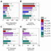
Mitochondrial respiration functional parameters were assessed using alterations in the ETC. (A) Spare respiratory capacity (%), which measures the difference between maximal respiration and basal respiration; (B) coupling efficiency (%) by measuring ATP production per available oxygen; (C) ATP production; and (D) proton leak across ETC. Statistical significance codes: treatment vs. untreated control (UT, *) and positive control (6-ODHA, +). Asterisks or plus signs indicate p-value ranges: *,+ 0.01–0.05, **,++ 0.001–0.01, ***,+++ 0.0001 to 0.001.
Figure 6
Figure 6
Principal component analyses by (A) treatment and (B) toxicological parameters, performed using R. Clustered treatments are more similar to variance in parameters and vice versa. The percent variance explained is provided for the two largest principal components. Untreated cells are labeled as UT.

Similar articles

Cited by

References

    1. Nakmode D.D., Day C.M., Song Y., Garg S. The Management of Parkinson’s Disease: An Overview of the Current Advancements in Drug Delivery Systems. Pharmaceutics. 2023;15:1503. doi: 10.3390/pharmaceutics15051503. - DOI - PMC - PubMed
    1. Dorsey E., Sherer T., Okun M., Bloem B. The emerging evidence of the Parkinson Pandemic. J. Park. Dis. 2018;8((Suppl. S1)):S3–S8. doi: 10.3233/JPD-181474. - DOI - PMC - PubMed
    1. Bloem B.R., Okun M.S., Klein C. Parkinson’s disease. Lancet. 2021;397:2284–2303. doi: 10.1016/S0140-6736(21)00218-X. - DOI - PubMed
    1. Ball N., Teo W.-P., Chandra S., Chapman J. Parkinson’s disease and the environment. Front. Neurol. 2019;10:218. doi: 10.3389/fneur.2019.00218. - DOI - PMC - PubMed
    1. Firestone J.A., Lundin J.I., Powers K.M., Smith-Weller T., Franklin G.M., Swanson P.D., Longstreth W., Jr., Checkoway H. Occupational factors and risk of Parkinson’s disease: A population-based case–control study. Am. J. Ind. Med. 2010;53:217–223. doi: 10.1002/ajim.20788. - DOI - PMC - PubMed