

Extracellular Vesicles and Their Role in Lung Infections
- PMID: 38003329
- PMCID: PMC10671184
- DOI: 10.3390/ijms242216139
Extracellular Vesicles and Their Role in Lung Infections
Abstract
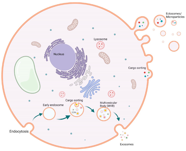
Lung infections are one of the most common causes of death and morbidity worldwide. Both bacterial and viral lung infections cause a vast number of infections with varying severities. Extracellular vesicles (EVs) produced by different cells due to infection in the lung have the ability to modify the immune system, leading to either better immune response or worsening of the disease. It has been shown that both bacteria and viruses have the ability to produce their EVs and stimulate the immune system for that. In this review, we investigate topics from EV biogenesis and types of EVs to lung bacterial and viral infections caused by various bacterial species. Mycobacterium tuberculosis, Staphylococcus aureus, and Streptococcus pneumoniae infections are covered intensively in this review. Moreover, various viral lung infections, including SARS-CoV-2 infections, have been depicted extensively. In this review, we focus on eukaryotic-cell-derived EVs as an important component of disease pathogenesis. Finally, this review holds high novelty in its findings and literature review. It represents the first time to cover all different information on immune-cell-derived EVs in both bacterial and viral lung infections.
Keywords: COVID-19; EVs; bacterial infection; biomarker; exosomes; extracellular vesicles; lung infection.
Conflict of interest statement
The authors declare no conflict of interest.
Figures
Similar articles
-
COVID-19 and Extracellular Vesicles: An Intriguing Interplay.Kidney Blood Press Res. 2020;45(5):661-670. doi: 10.1159/000511402. Epub 2020 Sep 21. Kidney Blood Press Res. 2020. PMID: 32957112 Free PMC article. Review.
-
Immunomodulatory Effects of Pneumococcal Extracellular Vesicles on Cellular and Humoral Host Defenses.mBio. 2018 Apr 10;9(2):e00559-18. doi: 10.1128/mBio.00559-18. mBio. 2018. PMID: 29636428 Free PMC article.
-
Proteomic profiling of single extracellular vesicles reveals colocalization of SARS-CoV-2 with a CD81/integrin-rich EV subpopulation in sputum from COVID-19 severe patients.Front Immunol. 2023 May 12;14:1052141. doi: 10.3389/fimmu.2023.1052141. eCollection 2023. Front Immunol. 2023. PMID: 37251406 Free PMC article.
-
Extracellular Vesicles in Viral Infections of the Nervous System.Viruses. 2020 Jun 28;12(7):700. doi: 10.3390/v12070700. Viruses. 2020. PMID: 32605316 Free PMC article. Review.
-
The Role of Extracellular Vesicles in Pandemic Viral Infections.J Microbiol. 2024 Jun;62(6):419-427. doi: 10.1007/s12275-024-00144-x. Epub 2024 Jun 25. J Microbiol. 2024. PMID: 38916789 Review.
Cited by
-
Stem cell-derived extracellular vesicles: novel therapeutics for cerebral injury following cardiac arrest and potential mechanisms.Cell Biosci. 2025 Jul 26;15(1):110. doi: 10.1186/s13578-025-01451-5. Cell Biosci. 2025. PMID: 40713691 Free PMC article. Review.
-
Exosomes: intriguing mediators of intercellular communication in the organism's response to noxious agents.Arh Hig Rada Toksikol. 2024 Dec 29;75(4):228-239. doi: 10.2478/aiht-2024-75-3923. eCollection 2024 Dec 1. Arh Hig Rada Toksikol. 2024. PMID: 39718095 Free PMC article. Review.
-
Particulate matter-induced epigenetic modifications and lung complications.Eur Respir Rev. 2024 Nov 13;33(174):240129. doi: 10.1183/16000617.0129-2024. Print 2024 Oct. Eur Respir Rev. 2024. PMID: 39537244 Free PMC article. Review.
References
Publication types
MeSH terms
LinkOut - more resources
Full Text Sources
Medical
Miscellaneous
