. 2023 Nov 10;24(22):16178.
doi: 10.3390/ijms242216178.

Dual RNA-Seq Analysis Pinpoints a Balanced Regulation between Symbiosis and Immunity in Medicago truncatula- Sinorhizobium meliloti Symbiotic Nodules

Affiliations

Dual RNA-Seq Analysis Pinpoints a Balanced Regulation between Symbiosis and Immunity in Medicago truncatula- Sinorhizobium meliloti Symbiotic Nodules

Dandan Zhang et al. Int J Mol Sci. .

Abstract

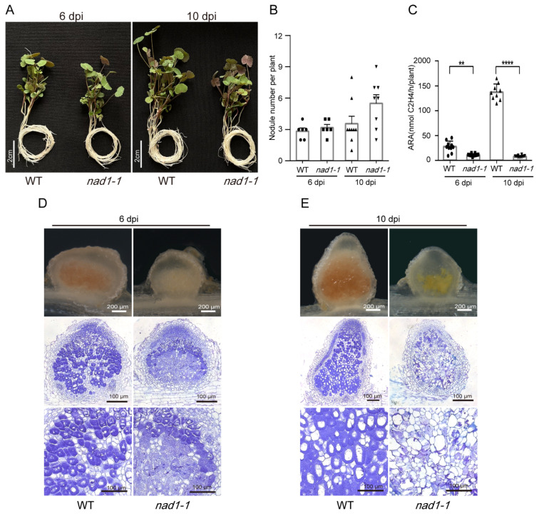
Legume-rhizobial symbiosis initiates the formation of root nodules, within which rhizobia reside and differentiate into bacteroids to convert nitrogen into ammonium, facilitating plant growth. This process raises a fundamental question: how is plant immunity modulated within nodules when exposed to a substantial number of foreign bacteria? In Medicago truncatula, a mutation in the NAD1 (Nodules with Activated Defense 1) gene exclusively results in the formation of necrotic nodules combined with activated immunity, underscoring the critical role of NAD1 in suppressing immunity within nodules. In this study, we employed a dual RNA-seq transcriptomic technology to comprehensively analyze gene expression from both hosts and symbionts in the nad1-1 mutant nodules at different developmental stages (6 dpi and 10 dpi). We identified 89 differentially expressed genes (DEGs) related to symbiotic nitrogen fixation and 89 DEGs from M. truncatula associated with immunity in the nad1-1 nodules. Concurrently, we identified 27 rhizobial DEGs in the fix and nif genes of Sinorhizobium meliloti. Furthermore, we identified 56 DEGs from S. meliloti that are related to stress responses to ROS and NO. Our analyses of nitrogen fixation-defective plant nad1-1 mutants with overactivated defenses suggest that the host employs plant immunity to regulate the substantial bacterial colonization in nodules. These findings shed light on the role of NAD1 in inhibiting the plant's immune response to maintain numerous rhizobial endosymbiosis in nodules.

Keywords: Medicago truncatula; NAD1; Sinorhizobium meliloti; dual RNA-seq; legume–rhizobial symbiosis; plant defense response.


Conflict of interest statement

The authors declare no conflict of interest.

Figures

Figure 1
Figure 1
Phenotypes of the wild-type (WT) Medicago truncatula R108 and nad1-1 mutant. (A) Growth of WT and nad1-1 at 6 days post-inoculation (dpi) and 10 dpi inoculated with Sinorhizobium meliloti 2011. (B) Nodule number per plant was measured at 6 dpi and 10 dpi. (C) Acetylene reduction assay (ARA) reflecting nitrogenase activity was performed on nodulated plants. **, p < 0.05; ****, p < 0.01. (D,E) Sections of WT and nad1-1 mutant nodules at 6 dpi and 10 dpi.
Figure 2
Figure 2
Identification of signature genes in WT and nad1-1 mutant. (A) Principal-component analysis (PCA) of the WT and nad1-1 transcriptome from two different times: nad1-1 6 dpi, wild type (WT) 6 dpi, nad1-1 10 dpi, wild type (WT) 10 dpi. (B) Number of differentially expressed genes (DEGs) in nad1-1 vs. WT at 6 dpi and 10 dpi. (C) Heatmap showing relative expression levels for the ‘‘nad1-1 signature’’, a set of 125 genes significantly regulated between nad1-1 vs. WT at 6 dpi, and 10 dpi in M. truncatula. (D) KEGG pathway enrichment analyses of the “nad1-1 signature” genes. (E) Violin plot showing expression levels of genes involved in the glutathione metabolism and oxidative phosphorylation pathways. (F) Heatmap showing relative expression levels for the genes in the other pathways (D). Gradient scale represents expression levels, with red showing the highest expression to blue with the lowest expression. “nad1-1 vs. WT” represents comparison of transcription of gene families in nad1-1 mutant nodules (nad1-1) vs. WT nodules (WT).
Figure 3
Figure 3
Analysis of different rhizobia transcriptional patterns in nad1-1 vs. WT. (A) Principal-component analysis (PCA) of the rhizobia transcriptome from two different times: nad1-1 6 dpi, wild type (WT) 6 dpi, nad1-1 10 dpi, wild type (WT) 10 dpi. (B) Number of rhizobia differentially expressed genes (DEGs) in nad1-1 vs. WT at 6 dpi and 10 dpi. (C) Heatmap showing the relative expression levels of rhizobial genes for the “Rhizobia signature”, a set of 25 genes significantly regulated in nad1-1 vs. WT at 6 dpi and 10 dpi. (D) KEGG pathway enrichment analyses of the “Rhizobia signature” genes. The red and blue dots represent p-values. (E) Heatmap showing expression levels of genes involved in the nitrogen metabolism, two-component system, and ABC transporter pathways. Gradient scale represents expression levels, with red showing the highest expression to blue with the lowest expression. “nad1-1 vs. WT” represents comparison of transcription of gene families in nad1-1 mutant nodules (nad1-1) vs. WT nodules (WT).
Figure 3
Figure 3
Analysis of different rhizobia transcriptional patterns in nad1-1 vs. WT. (A) Principal-component analysis (PCA) of the rhizobia transcriptome from two different times: nad1-1 6 dpi, wild type (WT) 6 dpi, nad1-1 10 dpi, wild type (WT) 10 dpi. (B) Number of rhizobia differentially expressed genes (DEGs) in nad1-1 vs. WT at 6 dpi and 10 dpi. (C) Heatmap showing the relative expression levels of rhizobial genes for the “Rhizobia signature”, a set of 25 genes significantly regulated in nad1-1 vs. WT at 6 dpi and 10 dpi. (D) KEGG pathway enrichment analyses of the “Rhizobia signature” genes. The red and blue dots represent p-values. (E) Heatmap showing expression levels of genes involved in the nitrogen metabolism, two-component system, and ABC transporter pathways. Gradient scale represents expression levels, with red showing the highest expression to blue with the lowest expression. “nad1-1 vs. WT” represents comparison of transcription of gene families in nad1-1 mutant nodules (nad1-1) vs. WT nodules (WT).
Figure 4
Figure 4
Candidate genes involved in the control of nod factor (NF) signaling. (A) Heatmap showing the transcriptional expression levels of all plant genes related to nodule NF signaling. Gradient scale represents expression levels, with red showing the highest expression to blue with the lowest expression. (B) Bar plot showing the expression levels of NF signaling-related genes regulated between nad1-1 and WT at 6 dpi and 10 dpi in M. truncatula. (C) Boxplot showing the expression levels of NF-genes regulated in nad1-1 vs. WT at 6 dpi and 10 dpi in S. meliloti. “nad1-1 vs. WT” represents comparison of transcription of gene families in nad1-1 mutant nodules (nad1-1) vs. WT nodules (WT).
Figure 5
Figure 5
Candidate genes involved in the control of the nodule meristem and differentiation. (A) Heatmap showing the transcriptional expression levels of all plant genes related to nodule meristem and differentiation. Gradient scale represents expression levels, with red showing the highest expression to blue with the lowest expression. (B,C) Gene ranking dot plots showing the DEGs related to nodule meristem and differentiation between nad1-1 and WT at 6 dpi (B) and 10 dpi (C) in M. truncatula. (D) Bar plot showing the genes related to nodule meristem and differentiation at 6 dpi and 10 dpi in S. meliloti. “nad1-1 vs. WT” represents comparison of transcription of gene families in nad1-1 mutant nodules (nad1-1) vs. WT nodules (WT).
Figure 6
Figure 6
Identification of plant and rhizobial transcriptional responses in symbiotic nitrogen fixation processes. Gradient scale represents expression levels, with red showing the highest expression to blue with the lowest expression. (A) Heatmap showing relative expression levels for the genes in symbiotic nitrogen fixation processes in M. truncatula. (B) Barblot showing expression profiles of leghemoglobin, NCR, and CaM-like genes in M. truncatula. (C) Dot chart plot showing expression profiles of nif and fix genes in S. meliloti. “nad1-1 vs. WT” represents comparison of transcription of gene families in nad1-1 mutant nodules (nad1-1) vs. WT nodules (WT).
Figure 7
Figure 7
Transcriptomic analysis of M. truncatula gene expression during defense. (A,B), Volcano plots showing differential expression of defense genes in nad1-1 vs. WT at 6 dpi (A) and 10 dpi (B). The top 10 genes ordered by log2 fold change are highlighted, and genes with an adjusted p-value of <0.05 are considered statistically significant. (C) Heatmap showing relative expression levels of defense-related genes in plant at 6 dpi and 10 dpi in nad1-1 vs. WT. Gradient scale represents expression levels, with red showing the highest expression to blue with the lowest expression. “nad1-1 vs. WT” represents comparison of transcription of gene families in nad1-1 mutant nodules (nad1-1) vs. WT nodules (WT).
Figure 8
Figure 8
Several pathways are regulated during defense in S. meliloti. (A) Expression profiles of defense-related genes at 6 dpi and 10 dpi in nad1-1 vs. WT. Gradient scale represents expression levels, with red showing the highest expression to blue with the lowest expression. (B) Boxplot of the main genes related to the plant-pathogen interaction pathway. (C) Violin plot showing expression levels of genes involved in the TCA cycle pathway. (D,F) Bar plots showing relative expression levels for genes in the bacterial secretion system (D), NO, ROS, and NCR pathways (F). (E) Bean plot showing expression levels of genes involved in the flagellar assembly. “nad1-1 vs. WT” represents comparison of transcription of gene families in nad1-1 mutant nodules (nad1-1) vs. WT nodules (WT).

Similar articles

Cited by

  • Defense and senescence interplay in legume nodules.
    Berrabah F, Benaceur F, Yin C, Xin D, Magne K, Garmier M, Gruber V, Ratet P. Berrabah F, et al. Plant Commun. 2024 Apr 8;5(4):100888. doi: 10.1016/j.xplc.2024.100888. Epub 2024 Mar 26. Plant Commun. 2024. PMID: 38532645 Free PMC article. Review.
  • CLE peptide signaling in plant-microbe interactions.
    Nakagami S, Kajiwara T, Tsuda K, Sawa S. Nakagami S, et al. Front Plant Sci. 2024 Oct 23;15:1481650. doi: 10.3389/fpls.2024.1481650. eCollection 2024. Front Plant Sci. 2024. PMID: 39507357 Free PMC article. Review.

References

    1. Oldroyd G.E.D., Leyser O. A plant’s diet, surviving in a variable nutrient environment. Science. 2020;368:eaba0196. doi: 10.1126/science.aba0196. - DOI - PubMed
    1. Mergaert P., Kereszt A., Kondorosi E. Gene Expression in Nitrogen-Fixing Symbiotic Nodule Cells in Medicago truncatula and Other Nodulating Plants. Plant Cell. 2020;32:42–68. doi: 10.1105/tpc.19.00494. - DOI - PMC - PubMed
    1. Wang Y.Y., Cheng Y.H., Chen K.E., Tsay Y.F. Nitrate Transport, Signaling, and Use Efficiency. Annu. Rev. Plant Biol. 2018;69:85–122. doi: 10.1146/annurev-arplant-042817-040056. - DOI - PubMed
    1. Guo K., Yang J., Yu N., Luo L., Wang E. Biological nitrogen fixation in cereal crops: Progress, strategies, and perspectives. Plant Commun. 2023;4:100499. doi: 10.1016/j.xplc.2022.100499. - DOI - PMC - PubMed
    1. Aasfar A., Bargaz A., Yaakoubi K., Hilali A., Bennis I., Zeroual Y., Meftah Kadmiri I. Nitrogen Fixing Azotobacter Species as Potential Soil Biological Enhancers for Crop Nutrition and Yield Stability. Front. Microbiol. 2021;12:628379. doi: 10.3389/fmicb.2021.628379. - DOI - PMC - PubMed

LinkOut - more resources