Review
. 2023 Nov 12;24(22):16233.
doi: 10.3390/ijms242216233.

Modulating Stress Proteins in Response to Therapeutic Interventions for Parkinson's Disease

Affiliations
Review

Modulating Stress Proteins in Response to Therapeutic Interventions for Parkinson's Disease

Serena Silvestro et al. Int J Mol Sci. .

Abstract

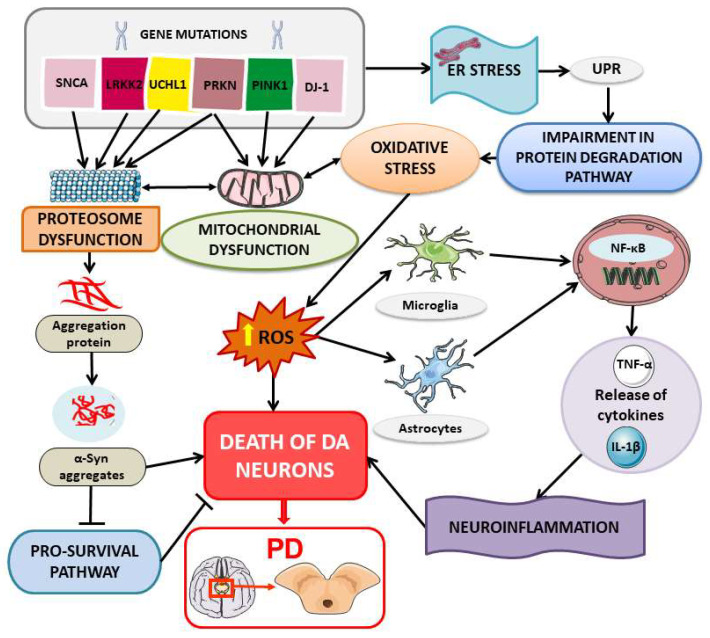
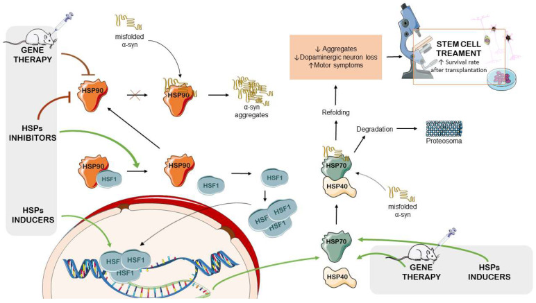
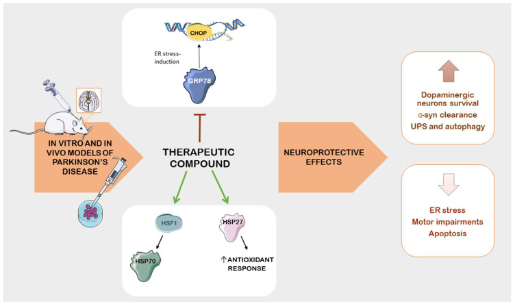
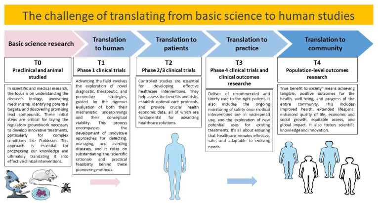
Parkinson's disease (PD) is a neurodegenerative illness characterized by the degeneration of dopaminergic neurons in the substantia nigra, resulting in motor symptoms and without debilitating motors. A hallmark of this condition is the accumulation of misfolded proteins, a phenomenon that drives disease progression. In this regard, heat shock proteins (HSPs) play a central role in the cellular response to stress, shielding cells from damage induced by protein aggregates and oxidative stress. As a result, researchers have become increasingly interested in modulating these proteins through pharmacological and non-pharmacological therapeutic interventions. This review aims to provide an overview of the preclinical experiments performed over the last decade in this research field. Specifically, it focuses on preclinical studies that center on the modulation of stress proteins for the treatment potential of PD. The findings display promise in targeting HSPs to ameliorate PD outcomes. Despite the complexity of HSPs and their co-chaperones, proteins such as HSP70, HSP27, HSP90, and glucose-regulated protein-78 (GRP78) may be efficacious in slowing or preventing disease progression. Nevertheless, clinical validation is essential to confirm the safety and effectiveness of these preclinical approaches.

Keywords: Parkinson’s disease; heat shock proteins; neuroregeneration; nonpharmacological interventions; pharmacological modulation; protein misfolding; stress protein modulation.


Conflict of interest statement

The authors declare no conflict of interest.

Figures

Figure 1
Figure 1
PRISMA flow diagram detailing the methodology that was applied to choose the preclinical studies that were used for the writing of the review. Duplicate articles were excluded from the total number of studies that were recorded. We selected the articles that described therapeutic strategies for improving PD symptoms and modulating stress proteins in preclinical studies. (The PRISMA Statement is published in [14]).
Figure 2
Figure 2
Heat shock proteins and their molecular mechanisms involved in PD pathogenesis. Stress proteins interact with several proteins involved in the processes of PD, such as α-syn and LRRK2. Mutations in PD-related genes can affect the interaction between these proteins and the HSP, altering the HSP’s ability to prevent protein aggregation or facilitate proper folding. Compromised HSPs may not effectively prevent α-syn aggregation, leading to the accumulation of toxic protein aggregates typical of the disease. Defective HSPs may not sustain the efficiency of the UPS, causing the accumulation of damaged proteins and promoting protein aggregation. Furthermore, altered HSPs may fail to mitigate oxidative stress, leaving cells vulnerable to damage caused by free radicals and reactive oxygen species (ROS). This could contribute to persistent inflammation, amplifying the inflammatory process that contributes to neuronal degeneration. Overall, dysfunctional HSPs could compromise cell survival, accelerating the death of neuronal cells in the context of PD pathogenesis. The image was created using the image bank of Servier Medical Art (available online: http://smart.servier.com/, accessed on 15 September 2023), licensed under a Creative Commons Attribution 3.0 Unported License (available online: https://creativecommons.org/licenses/by/3.0/, accessed on 15 September 2023). PD: Parkinson’s disease; SNCA: synuclein alpha; LRRK2: leucine-rich repeat kinase 2; UCHL1: ubiquitin C-terminal hydrolase L1; PRKN: parkin; PINK1: phosphatase and tensin homolog-induced kinase 1; α-Syn: alpha synuclein; ER: endoplasmic reticulum; UPR: unfolded protein response; ROS: reactive oxygen species; NF-κB: nuclear factor kappa-light-chain-enhancer of activated B cells; TNF-α: tumor necrosis factor alpha; IL-1β: interleukin-1β.
Figure 3
Figure 3
The main therapeutic approaches targeting the modulation of stress proteins to reduce the impact of PD. The simultaneous employment of pharmacological and genetic strategies and stem cell therapies may offer the possibility of protecting neuronal cells, thus preventing the principal etiopathogenesis processes of PD. The image was created using the image bank of Servier Medical Art (available online: http://smart.servier.com/, accessed on 15 September 2023), licensed under a Creative Commons Attribution 3.0 Unported License (available online: https://creativecommons.org/licenses/by/3.0/, accessed on 15 September 2023). HSPs: heat shock proteins; HSF-1: heat shock factor-1; α-syn: alpha-synuclein.
Figure 4
Figure 4
The main HSPs modulated by therapeutic compounds in preclinical models of PD. Therapies, both chemical and natural, regulate pathological changes in PD by targeting specific chaperone proteins and reducing ER stress. Key chaperones involved include GPR78/CHOP, HSF1/HSP70, and HSP27. Modulation of HSPs by therapeutic compounds preserves dopaminergic neurons, enhances α-syn degradation, activates the UPS, and promotes autophagy. Simultaneously, this neuroprotective effect leads to the suppression of ER stress, alleviation of motor deficits, and a decrease in cell death. The image was created using the image bank of Servier Medical Art (available online: http://smart.servier.com/, accessed on 15 September 2023), licensed under a Creative Commons Attribution 3.0 Unported License (available online: https://creativecommons.org/licenses/by/3.0/, accessed on 15 September 2023). HSPs: heat shock proteins; PD: Parkinson’s disease; ER: endoplasmic reticulum; GPR78: G protein-coupled receptor 78; CHOP: C/EBP homologous protein; HSF-1: heat shock factor-1; α-syn: alpha synuclein; UPS: ubiquitin–proteasome system.
Figure 5
Figure 5
Translational research involves several operational phases (T0-T4) and the associated challenges that must be addressed. T0 represents basic science research focused on understanding cellular mechanisms, their links to diseases, and the identification of potential therapeutic targets, along with the development of new treatment methods, such as new drugs. T1 encompasses proof-of-concept studies conducted in human volunteers during phase 1 clinical trials. These studies aim to establish the safety, mechanisms, and feasibility of the proposed concept. T2 involves phase 2 and 3 clinical trials, ideally randomized, to test the effectiveness of the therapeutic agent in patient populations that represent the specific disease under consideration. These trials often include control groups for comparison. T3 is associated with phase 4 clinical trials, which aim to optimize the practical use of a therapeutic agent in clinical settings. T4 refers to population-level outcomes research or comparative effectiveness research, which assesses the long-term utility and cost-effectiveness of a therapeutic agent in comparison with other treatments in current use. Translating findings from basic science to human studies is a critical and challenging process. The image was created using the image bank of Servier Medical Art (available online: http://smart.servier.com/, accessed on 30 October 2023), licensed under a Creative Commons Attribution 3.0 Unported License (available online: https://creativecommons.org/licenses/by/3.0/, accessed on 30 October 2023).

Similar articles

Cited by

References

    1. Ascherio A., Schwarzschild M.A. The epidemiology of parkinson’s disease: Risk factors and prevention. Lancet Neurol. 2016;15:1257–1272. doi: 10.1016/S1474-4422(16)30230-7. - DOI - PubMed
    1. Schneider S.A., Obeso J.A. Clinical and pathological features of parkinson’s disease. Behav. Neurobiol. Huntington’s Dis. Park. Dis. 2015;22:205–220. - PubMed
    1. Sveinbjornsdottir S. The clinical symptoms of parkinson’s disease. J. Neurochem. 2016;139((Suppl. 1)):318–324. doi: 10.1111/jnc.13691. - DOI - PubMed
    1. Kudryavtseva A.V., Krasnov G.S., Dmitriev A.A., Alekseev B.Y., Kardymon O.L., Sadritdinova A.F., Fedorova M.S., Pokrovsky A.V., Melnikova N.V., Kaprin A.D., et al. Mitochondrial dysfunction and oxidative stress in aging and cancer. Oncotarget. 2016;7:44879–44905. doi: 10.18632/oncotarget.9821. - DOI - PMC - PubMed
    1. Jellinger K.A. Neuropathology and pathogenesis of extrapyramidal movement disorders: A critical update—I. Hypokinetic-rigid movement disorders. J. Neural Transm. 2019;126:933–995. doi: 10.1007/s00702-019-02028-6. - DOI - PubMed

MeSH terms

Substances