. 2023 Nov 14;24(22):16284.
doi: 10.3390/ijms242216284.

Epidermal Growth Factor Receptors in Vascular Endothelial Cells Contribute to Functional Hyperemia in the Brain

Affiliations

Epidermal Growth Factor Receptors in Vascular Endothelial Cells Contribute to Functional Hyperemia in the Brain

Hannah R Ferris et al. Int J Mol Sci. .

Abstract

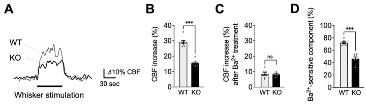
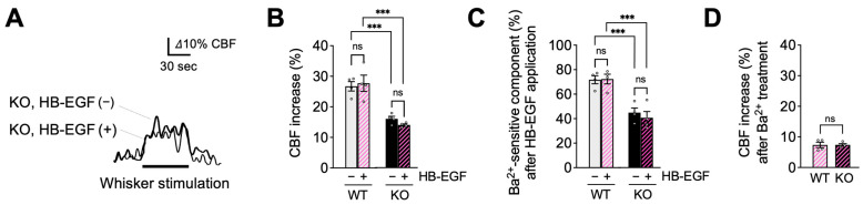
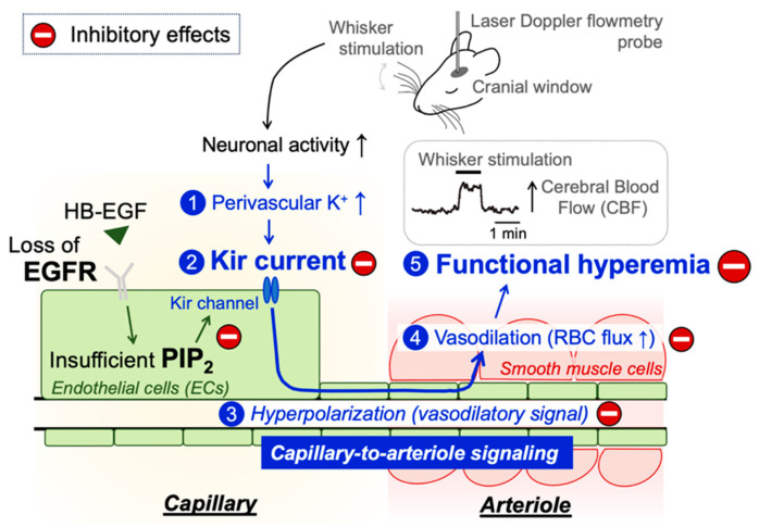
Functional hyperemia-activity-dependent increases in local blood perfusion-underlies the on-demand delivery of blood to regions of enhanced neuronal activity, a process that is crucial for brain health. Importantly, functional hyperemia deficits have been linked to multiple dementia risk factors, including aging, chronic hypertension, and cerebral small vessel disease (cSVD). We previously reported crippled functional hyperemia in a mouse model of genetic cSVD that was likely caused by depletion of phosphatidylinositol 4,5-bisphosphate (PIP2) in capillary endothelial cells (EC) downstream of impaired epidermal growth factor receptor (EGFR) signaling. Here, using EC-specific EGFR-knockout (KO) mice, we directly examined the role of endothelial EGFR signaling in functional hyperemia, assessed by measuring increases in cerebral blood flow in response to contralateral whisker stimulation using laser Doppler flowmetry. Molecular characterizations showed that EGFR expression was dramatically decreased in freshly isolated capillaries from EC-EGFR-KO mice, as expected. Notably, whisker stimulation-induced functional hyperemia was significantly impaired in these mice, an effect that was rescued by administration of PIP2, but not by the EGFR ligand, HB-EGF. These data suggest that the deletion of the EGFR specifically in ECs attenuates functional hyperemia, likely via depleting PIP2 and subsequently incapacitating Kir2.1 channel functionality in capillary ECs. Thus, our study underscores the role of endothelial EGFR signaling in functional hyperemia of the brain.

Keywords: cerebral small vessel diseases (cSVD); epidermal growth factor receptor (EGFR); functional hyperemia; phosphatidylinositol 4,5-bisphosphate (PIP2); vascular endothelial cells.


Conflict of interest statement

The authors declare no conflict of interest.

Figures

Figure 1
Figure 1
Characterization of capillary ECs from EC-EGFR-KO mice. (A,B): Bright field and fluorescent images of isolated microvessels from EC/SMC dual-reporter mice before (A) and after two-step filter purification (B). Green fluorescent protein expresses in ECs under the cadherin-5 (cdh5) promotor, and red fluorescent protein expresses in SMC/pericytes under the acta2 promoter. The vast majority (>98%, counted by cell numbers after DAPI staining) of tissue obtained by two-step filter purification was green fluorescence-positive and red fluorescence-negative microvessels, demonstrating that the collected tissue samples were capillaries. White arrows indicate microvessels in filter-purified microvessel fraction (panel B). In contrast, the crude microvessel fraction (before the two-step filter-purification) contains red fluorescence-positive microvessels (i.e., arterioles). Note: The green fluorescent image of crude microvessel fraction also shows auto-fluorescence in elastic lamina membranes and brain tissue debris. An example of green fluorescence-positive and red fluorescence-negative microvessels (i.e., capillaries) in crude microvessel fractions are circled by white dotted lines. Experiments were repeated four times and exhibited consistent and similar results. (C): EGFR protein in freshly isolated capillaries, pooled from 4 animals for each sample, were quantified by ELISA. EGFR protein was dramatically decreased in EC-EGFR-KO mice compared to WT animals (i.e., Cre-negative littermates). Data are presented as mean ± SEM (n = 8 samples from 32 WT animals, n = 6 samples from 24 KO mice). *** p < 0.001 between groups by unpaired t-test.
Figure 2
Figure 2
Impaired functional hyperemia in EC-EGFR-KO mice. (A): Representative traces showing CBF increases in contralateral somatosensory cortex during whisker stimulation. Cre-negative littermates (WT) were used as a control group in comparison to EC-EGFR-KO (KO) mice. (B): Summary data of whisker stimulation-induced functional hyperemia. (C): CBF increase during whisker stimulation after treatment with Ba2+ (100 μM), a Kir2.1 channel blocker. (D): Ba2+-sensitive component of functional hyperemia, i.e., the difference in responses before and after Ba2+ treatment, indicating the contribution of the Kir2.1 channels to whisker stimulation-induced functional hyperemia. Data are presented as mean ± SEM (n = 8 animals in WT, n = 8 animals in KO). *** p < 0.001, ns: not significant between groups by unpaired t-test.
Figure 3
Figure 3
PIP2, an endogenous Kir2.1 channel co-factor, restored functional hyperemia in EC-EGFR-KO mice. (A): Whisker stimulation-induced functional hyperemia before and after PIP2 treatment in EC-EGFR-KO mice. (B): Summary data showing PIP2 treatment restores functional hyperemia deficits in EC-EGFR-KO mice, with little impact on WT animals. (C): Ba2+-sensitive component of functional hyperemia before and after PIP2 treatment. (D): CBF increase during whisker stimulation after Ba2+ treatment. Ba2+ was cortically superfused for 20 min following PIP2 treatment. Data are presented as mean ± SEM (n = 4 animals in WT, n = 4 animals in KO). *** p < 0.001, ** p < 0.01, * p < 0.05, ns: not significant between groups by two-way ANOVA (B,C) or by unpaired t-test (D).
Figure 4
Figure 4
HB-EGF, an EGFR ligand, failed to restore functional hyperemia in EC-EGFR-KO mice. (A): Whisker stimulation-induced functional hyperemia before and after HB-EGF treatment in EC-EGFR-KO mice. (B): Summary data showing that HB-EGF treatment did not alter whisker stimulation-induced functional hyperemia in either EC-EGFR-KO or WT mice. (C): Ba2+-sensitive component of functional hyperemia before and after HB-EGF treatment. (D): Whisker stimulation-induced CBF increase after Ba2+ treatment. Ba2+ was subsequently and concurrently applied for 20 min by adding to HB-EGF-containing cortical superfusate. Data are presented as mean ± SEM (n = 4 animals in WT, n = 4 animals in KO). *** p < 0.001, ns: not significant between groups by two-way ANOVA (B,C) or by unpaired t-test (C).
Figure 5
Figure 5
Schematic illustration of the proposed signaling pathway underlying functional hyperemia deficits in EC-EGFR-KO mice. Whisker stimulation and subsequent neuronal activation, which increases perivascular K+ during neuronal repolarization, results in Kir2.1 channel activation in capillary ECs. Kir2.1 channel-initiated hyperpolarizing vasodilatory signal rapidly propagates upstream and dilates the precapillary arteriole, increasing downstream tissue perfusion. This local increase in blood flow in neuronally active regions of the brain is termed functional hyperemia, which is detected using laser Doppler flowmetry in this study. Genetic deletion of EGFR disrupts functional hyperemia in response to whisker stimulation by causing Kir2.1 channel dysfunction, which is likely attributed to lowering the level of PIP2, an essential co-factor of Kir2.1 channels.

Update of

Similar articles

Cited by

References

    1. Iadecola C. The Neurovascular Unit Coming of Age: A Journey through Neurovascular Coupling in Health and Disease. Neuron. 2017;96:17–42. doi: 10.1016/j.neuron.2017.07.030. - DOI - PMC - PubMed
    1. Schaeffer S., Iadecola C. Revisiting the neurovascular unit. Nat. Neurosci. 2021;24:1198–1209. doi: 10.1038/s41593-021-00904-7. - DOI - PMC - PubMed
    1. Lecrux C., Hamel E. The neurovascular unit in brain function and disease. Acta Physiol. 2011;203:47–59. doi: 10.1111/j.1748-1716.2011.02256.x. - DOI - PubMed
    1. Santisteban M.M., Iadecola C., Carnevale D. Hypertension, Neurovascular Dysfunction, and Cognitive Impairment. Hypertension. 2023;80:22–34. doi: 10.1161/HYPERTENSIONAHA.122.18085. - DOI - PMC - PubMed
    1. Haydon P.G., Carmignoto G. Astrocyte control of synaptic transmission and neurovascular coupling. Physiol. Rev. 2006;86:1009–1031. doi: 10.1152/physrev.00049.2005. - DOI - PubMed

LinkOut - more resources