. 2023 Nov 15;24(22):16342.
doi: 10.3390/ijms242216342.

Tff3 Deficiency Differentially Affects the Morphology of Male and Female Intestines in a Long-Term High-Fat-Diet-Fed Mouse Model

Affiliations

Tff3 Deficiency Differentially Affects the Morphology of Male and Female Intestines in a Long-Term High-Fat-Diet-Fed Mouse Model

Kate Šešelja et al. Int J Mol Sci. .

Abstract

Trefoil factor family protein 3 (Tff3) protects the gastrointestinal mucosa and has a complex mode of action in different tissues. Here, we aimed to determine the effect of Tff3 deficiency on intestinal tissues in a long-term high-fat-diet (HFD)-fed model. A novel congenic strain without additional metabolically relevant mutations (Tff3-/-/C57Bl6NCrl strain, male and female) was used. Wild type (Wt) and Tff3-deficient mice of both sexes were fed a HFD for 36 weeks. Long-term feeding of a HFD induces different effects on the intestinal structure of Tff3-deficient male and female mice. For the first time, we found sex-specific differences in duodenal morphology. HFD feeding reduced microvilli height in Tff3-deficient females compared to that in Wt females, suggesting a possible effect on microvillar actin filament dynamics. These changes could not be attributed to genes involved in ER and oxidative stress, apoptosis, or inflammation. Tff3-deficient males exhibited a reduced cecal crypt depth compared to that of Wt males, but this was not the case in females. Microbiome-related short-chain fatty acid content was not affected by Tff3 deficiency in HFD-fed male or female mice. Sex-related differences due to Tff3 deficiency imply the need to consider both sexes in future studies on the role of Tff in intestinal function.

Keywords: ER stress; apoptosis; cecum; duodenum; high-fat diet; inflammation; oxidative stress; trefoil peptide 3.


Conflict of interest statement

The authors declare no conflict of interest.

Figures

Figure 1
Figure 1
Effect of long-term HFD feeding on body weight and blood glucose levels in Wt and Tff3-deficient mice of both sexes (n~10 animals per group). (a) Bodyweights of Wt and Tff3-/- mice (male and female) and blood glucose levels (b) at the age of 40 weeks, after 36 weeks of HFD. ** p ≤ 0.01.
Figure 2
Figure 2
Photomicrographs of the duodenum and cecum. Villus height and crypt depth were measured as indicated. In the duodenum, villus height was measured from the tip of the villus to the crypt–villus junction, and crypt depth was measured from the crypt–villus junction to its base. Measurement of crypt depth is shown in the cecum. H&E staining; scale bars 200 µm (duodenum) and 100 µm (cecum).
Figure 3
Figure 3
Duodenal ultrastructure of Wt and Tff3-/- mice fed a HFD. (a) Wt male; (b) Tff3-/- male; (c) Wt female; (d) Tff3-/- female. The height of the microvilli in the duodenum of Tff3-/- female mice is reduced (→); scale bars are 20 µm and 1 µm (inserts).
Figure 4
Figure 4
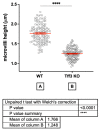
Height of microvilli in the duodenum of Wt and Tff3-/- female mice fed HFD. The microvilli in the duodenum of Tff3-/- female mice were significantly reduced. Compared with Wt females, Tff3-deficient females had 30% lower microvilli height. We analyzed 3 animals per group, and 4 different sections were presented for each animal. In each section, the height of at least 10 different microvilli was measured, red line is interval bar of SEM, **** p < 0.0001. Data were analyzed using ImageJ software (Version 1.50i, National Institute of Health, Bethesda, Md, USA) and t-test analysis.
Figure 5
Figure 5
Effect of long-term HFD feeding on the expression of ER stress markers in the duodenum of Wt and Tff3-deficient mice of both sexes. We performed qPCR using SYBR green detection for each group (n = 5 animals per group). Ct data were analyzed by the REST program and are presented as fold change with upper limits. (a) The expression levels of genes in Tff3-/- male mice relative to those of the Wt male mice are shown; (b) The expression levels of genes in Tff3-/- female mice relative to those of the Wt female mice are shown; (c) Expression levels of genes in Wt female mice relative to those of the Wt male mice are shown; (d) The expression of genes in Tff3-/- female mice relative to that of the Tff3-/- male mice is shown. * p ≤ 0.05.
Figure 6
Figure 6
Effect of long-term HFD feeding on the expression of oxidative stress markers in the duodenum of Wt and Tff3-deficient mice of both sexes. We performed qPCR using SYBR green detection for each group (n = 5 animals per group). Ct data were analyzed by the REST program and are presented as fold change with upper limits. (a) The expression levels of genes in Tff3-/- male mice relative to those of the Wt male mice are shown; (b) The expression levels of genes in Tff3-/- female mice relative to those of the Wt female mice are shown; (c) Expression levels of genes in Wt female mice relative to those of the Wt male mice are shown; (d) The expression levels of genes in Tff3-/- female mice relative to those of the Tff3-/- male mice are shown.
Figure 7
Figure 7
Effect of long-term HFD feeding on the expression of inflammation-relevant genes in the duodenum of Wt and Tff3-deficient mice of both sexes. We performed qPCR using SYBR green detection for each group (n = 5 animals per group). Ct data were analyzed by the REST program and are presented as fold change with upper limits. (a) Wt male mice; (b) The expression levels of genes in Tff3-/- female mice relative to those of the Wt female mice are shown; (c) Expression levels of genes in Wt female mice relative to those of the Wt male mice are shown; (d) The expression levels of genes in Tff3-/- female mice relative to those of the Tff3-/- male mice are shown.
Figure 8
Figure 8
Effect of long-term high-fat diet treatment on the expression of apoptosis-related genes in the duodenum of Wt and Tff3-deficient mice of both sexes. We performed qPCR using SYBR green detection for each group (n = 5 animals per group). Ct data were analyzed by the REST program and are presented as fold change with upper limits. (a) The expression levels of genes in Tff3-/- male mice relative to those of the Wt male mice are shown; (b) The expression levels of genes in Tff3-/- female mice relative to those of the Wt female mice are shown; (c) Expression levels of genes in Wt female mice relative to those of the Wt male mice are shown; (d) The expression levels of genes in Tff3-/- female mice relative to those of the Tff3-/- male mice are shown.

Similar articles

Cited by

References

    1. Thim L., May F.E.B. Structure of mammalian trefoil factors and functional insights. Cell. Mol. Life Sci. 2005;62:2956–2973. doi: 10.1007/s00018-005-5484-6. - DOI - PMC - PubMed
    1. Braga Emidio N., Brierley S.M., Schroeder C.I., Muttenthaler M. Structure, Function, and Therapeutic Potential of the Trefoil Factor Family in the Gastrointestinal Tract. ACS Pharmacol. Transl. Sci. 2020;3:583–597. doi: 10.1021/acsptsci.0c00023. - DOI - PMC - PubMed
    1. Hoffman W., Jagla W., Wiede A. Molecular medicine of TFF-peptides: From gut to brain. Histol. Histopathol. 2001;16:319–334. doi: 10.14670/HH-16.319. - DOI - PubMed
    1. Madsen J., Nielsen O., Tornøe I., Thim L., Holmskov U. Tissue localization of human trefoil factors 1, 2, and 3. J. Histochem. Cytochem. 2007;55:505–513. doi: 10.1369/jhc.6A7100.2007. - DOI - PubMed
    1. Baus-Loncar M., Schmid J., Lalani E., Goodlad R.A., Stamp G.W.H., Blin N. Trefoil Factor 2 (Tff2) Deficiency in Murine Digestive Tract Influences the Immune System. Cell Physiol. Biochem. 2005;16:31–42. doi: 10.1159/000087729. - DOI - PubMed