. 2023 Nov 16;24(22):16394.
doi: 10.3390/ijms242216394.

SARS-CoV-2 Spike Protein Intensifies Cerebrovascular Complications in Diabetic hACE2 Mice through RAAS and TLR Signaling Activation

Affiliations

SARS-CoV-2 Spike Protein Intensifies Cerebrovascular Complications in Diabetic hACE2 Mice through RAAS and TLR Signaling Activation

Faith N Burnett et al. Int J Mol Sci. .

Abstract

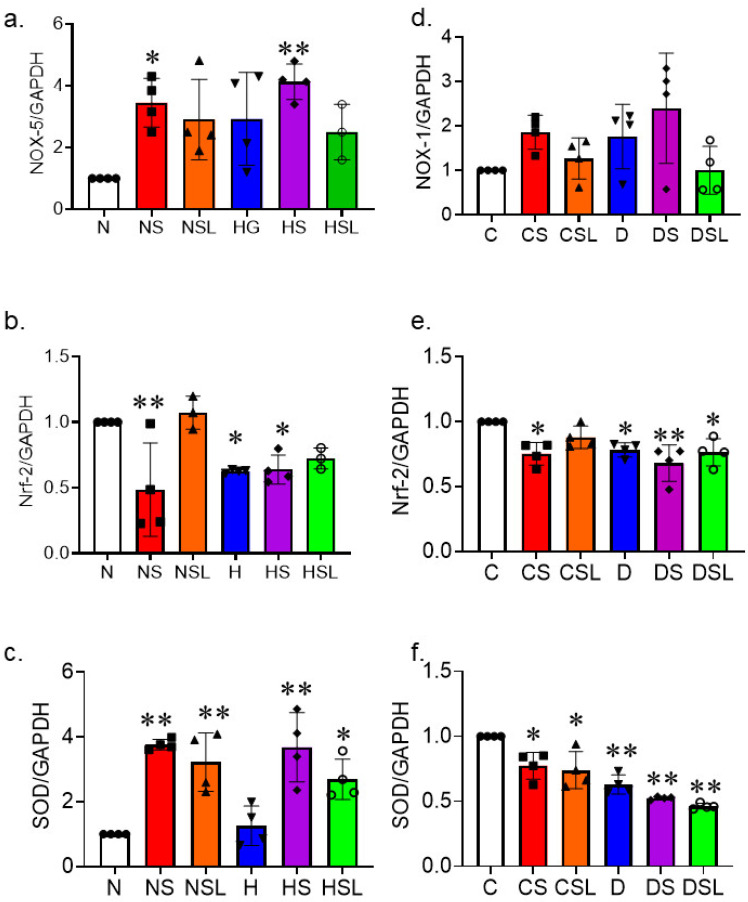
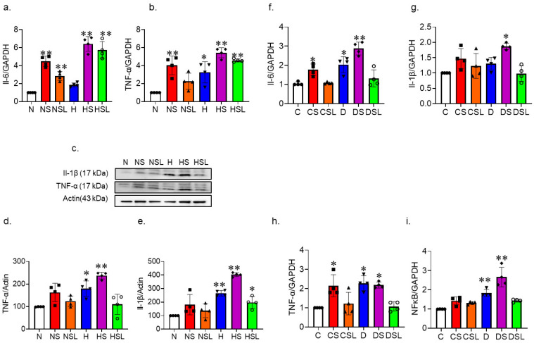
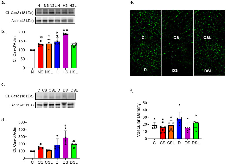
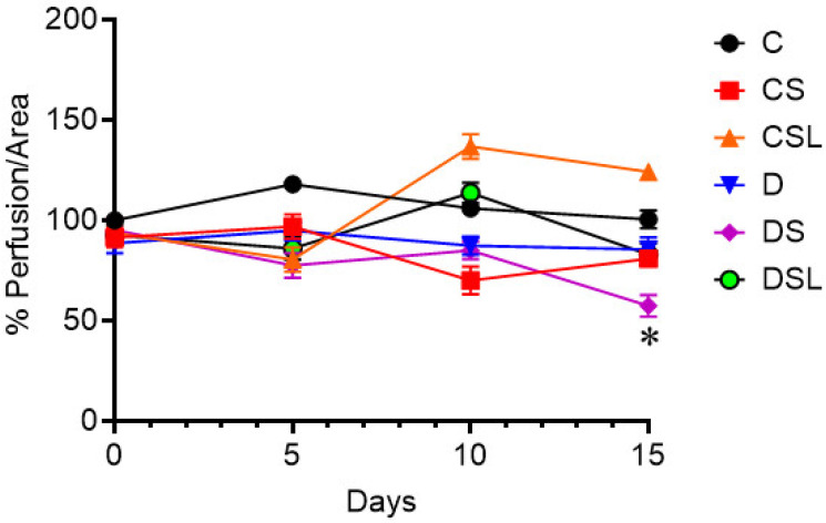
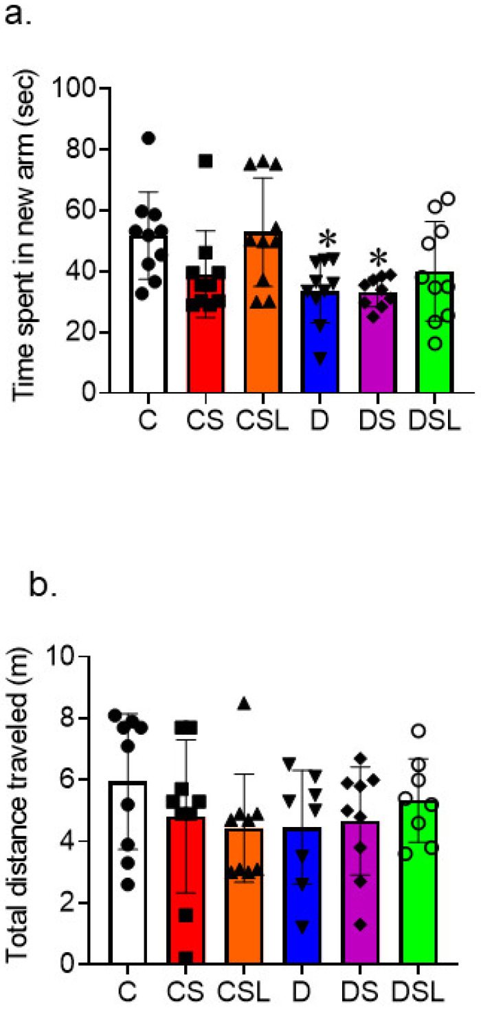
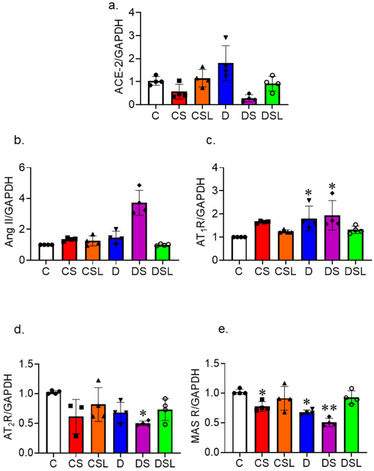
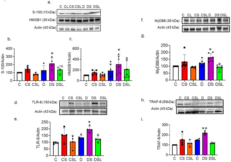
Diabetics are more vulnerable to SARS-CoV-2 neurological manifestations. The molecular mechanisms of SARS-CoV-2-induced cerebrovascular dysfunction in diabetes are unclear. We hypothesize that SARS-CoV-2 exacerbates diabetes-induced cerebrovascular oxidative stress and inflammation via activation of the destructive arm of the renin-angiotensin-aldosterone system (RAAS) and Toll-like receptor (TLR) signaling. SARS-CoV-2 spike protein was injected in humanized ACE2 transgenic knock-in mice. Cognitive functions, cerebral blood flow, cerebrovascular architecture, RAAS, and TLR signaling were used to determine the effect of SARS-CoV-2 spike protein in diabetes. Studies were mirrored in vitro using human brain microvascular endothelial cells treated with high glucose-conditioned media to mimic diabetic conditions. Spike protein exacerbated diabetes-induced cerebrovascular oxidative stress, inflammation, and endothelial cell death resulting in an increase in vascular rarefaction and diminished cerebral blood flow. SARS-CoV-2 spike protein worsened cognitive dysfunction in diabetes compared to control mice. Spike protein enhanced the destructive RAAS arm at the expense of the RAAS protective arm. In parallel, spike protein significantly exacerbated TLR signaling in diabetes, aggravating inflammation and cellular apoptosis vicious circle. Our study illustrated that SAR-CoV-2 spike protein intensified RAAS and TLR signaling in diabetes, increasing cerebrovascular damage and cognitive dysfunction.

Keywords: RAAS; SARS-CoV-2 spike protein; TLR signaling; cerebrovasculature; diabetes; hACE2 KI mice.


Conflict of interest statement

The authors declare no conflict of interest.

Figures

Figure 1
Figure 1
SARS-CoV-2 spike protein exacerbated diabetes-induced oxidative stress. Human brain microvascular endothelial cells were grown to confluency in complete media. Cells were then treated with SARS-CoV-2 spike protein and losartan under either normal glucose (complete media with equimolar L-glucose) or high glucose (D-glucose (25 mM) and sodium palmitate (200 µM)) conditions. (N, closed circles) normal glucose, (NS, closed squares) normal glucose + spike protein, (NSL, closed triangles) normal glucose + spike protein + losartan, (H, inverted triangles) high glucose, (HS, closed diamonds) high glucose + spike protein, (HSL, open circles) high glucose + spike protein + losartan. RNA was isolated and used for qRT-PCR assessment of oxidative stress. (a) RT-PCR analysis results show that spike protein exacerbated increases in NOX-5 caused by high glucose conditions. Losartan reduced the increases in NOX-5 gene expression following S-protein exposure under high glucose conditions (one-way ANOVA, * p = 0.02, ** p = 0.002, n = 4). (b) RT-PCR analysis results show that spike protein decreases cellular antioxidant defense by reducing Nrf-2 gene expression (one-way ANOVA, ** p = 0.001, * p = 0.018, n = 4). (c) RT-PCR analysis results show that spike protein decreases SOD gene expression (one-way ANOVA, ** p < 0.001, ** p = 0.002, n = 4). Type 2 diabetes was induced in hACE2 mice with low-dose STZ (35 mg/kg), followed by a high-fat diet (45 kcal% fat) for eight weeks. Mice were intravenously injected with SARS-CoV-2 spike protein (4 µg/animal) and treated with losartan (10 mg/kg body weight). (C, closed circles) control hACE2, (CS, closed squares) hACE2 + spike protein, (CSL, closed triangles) hACE2 + spike protein + losartan, (D, inverted triangles) diabetic hACE2, (DS, closed diamond) diabetic hACE2 + spike protein, (DSL, open circles) diabetic hACE2 + spike protein + losartan. RNA was isolated from whole brain lysate and used for qRT-PCR assessment of oxidative stress. (d) RT-PCR analysis results for NOX-1 gene expression (one-way ANOVA, p < 0.053, n = 4). (e) RT-PCR analysis for Nrf-2 gene expression in hACE2 mice (one-way ANOVA, * p = 0.011, ** p = 0.001, n = 4). (f) RT-PCR analysis for SOD expression in hACE2 mice (one-way ANOVA, * p = 0.008, ** p = 0.001, n = 4).
Figure 2
Figure 2
SARS-CoV-2 spike protein aggravates diabetes-induced inflammation. Human brain microvascular endothelial cells were grown to confluency in complete media. Cells were then treated with SARS-CoV-2 spike protein and losartan under either normal glucose (complete media with equimolar L-glucose) or high glucose (D-glucose (25 mM) and sodium palmitate (200 µM)) conditions. (N, closed circles) normal glucose, (NS, closed squares) normal glucose + spike protein, (NSL, closed triangles) normal glucose + spike protein + losartan, (H, inverted triangles) high glucose, (HS, closed diamonds) high glucose + spike protein, (HSL, open circles) high glucose + spike protein + losartan. RNA and protein were isolated from cell lysate and used for qRT-PCR and Western blot assessment of inflammation. (a) RT-PCR analysis showing the effect of spike protein on Il-6 gene expression (one-way ANOVA, ** p = 0.001, n = 4). (b) RT-PCR analysis showing effect of spike protein on TNF-α gene expression (one-way ANOVA, * p = 0.007, ** p = 0.001, n = 4). (c) Western blot representative of TNF-α and Il-1β. (d) Western blot analysis for TNF-α (one-way ANOVA, * p = 0.017, ** p = 0.001, n = 4). (e) Western blot analysis for Il-1β (one-way ANOVA, ** p = 0.001, * p = 0.045, n = 4). Type 2 diabetes was induced in hACE2 mice with low-dose STZ (35 mg/kg), followed by a high-fat diet (45 kcal% fat) for eight weeks. Mice were intravenously injected with SARS-CoV-2 spike protein (4 µg/animal) and treated with losartan (10 mg/kg body weight). (C, closed circles) control hACE2, (CS, closed squares) hACE2 + spike protein, (CSL, closed triangles) hACE2 + spike protein + losartan, (D, inverted triangles) diabetic hACE2, (DS, closed diamonds) diabetic hACE2 + spike protein, (DSL, open circles) diabetic hACE2 + spike protein + losartan. RNA was isolated from whole brain lysate and used for qRT-PCR assessment of inflammatory markers. We used qRT-PCR analysis for Il-6, Il-1β, TNF-α, and NFκB expression in whole brain homogenate. (f) RT-PCR analysis for Il-6 gene expression (one-way ANOVA, * p = 0.042, ** p = 0.001, n = 4). (g) RT-PCR analysis for Il-1β gene expression (one-way ANOVA, * p = 0.002, n = 4). (h) RT-PCR analysis for TNF-α gene expression (one-way ANOVA, * p = 0.006, n = 4). (i) RT-PCR analysis for NFκB gene expression (one-way ANOVA, ** p = 0.001, n = 4).
Figure 3
Figure 3
SARS-CoV-2 spike protein increases diabetes-induced endothelial cell death and vascular rarefaction. Human brain microvascular endothelial cells were grown to confluency in complete media. Cells were then treated with SARS-CoV-2 spike protein and losartan under either normal glucose (complete media with equimolar L-glucose) or high glucose (D-glucose (25 mM) and sodium palmitate (200 µM)) conditions. (N, closed circles) normal glucose, (NS, closed squares) normal glucose + spike protein, (NSL, closed triangles) normal glucose + spike protein + losartan, (H, inverted triangles) high glucose, (HS, closed diamonds) high glucose + spike protein, (HSL, open circles) high glucose + spike protein + losartan. Cell lysate was used for Western blot for assessment of endothelial cell death. (a,b) Western blot representative and quantification for cleaved caspase-3 protein expression (One-way ANOVA, * p = 0.021, ** p = 0.001, n = 4). (c,d) Type 2 diabetes was induced in hACE2 mice with low-dose STZ (35 mg/kg), followed by a high-fat diet (45 kcal% fat) for eight weeks. Mice were intravenously injected with SARS-CoV-2 spike protein (4 µg/animal) and treated with losartan (10 mg/kg body weight). (C, closed circles) control hACE2, (CS, closed squares) hACE2 + spike protein, (CSL, closed triangles) hACE2 + spike protein + losartan, (D, inverted triangles) diabetic hACE2, (DS, closed diamonds) diabetic hACE2 + spike protein, (DSL, open circles) diabetic hACE2 + spike protein + losartan. Whole brain homogenate was used to assess apoptosis using immunoblotting (one-way ANOVA, * p = 0.002, n = 4). (e,f) Vascular cell death and rarefaction was assessed in brain using immunohistochemical staining. Brains were sectioned and stained with Lycopersicon esculentum lectin, DyLight™ 488 to analyze cerebrovascular architecture. Diabetes alone increased vascular density compared to control hACE2 mice. SARS-CoV-2 spike protein caused a decrease in vascular density in both control and diabetic mice, but the effects were much more prominent in the diabetic group (20× magnification, one-way ANOVA, p = 0.014, n = 6–7).
Figure 4
Figure 4
SARS-CoV-2 spike protein exacerbate cerebral blood flow in diabetics. Type 2 diabetes was induced in hACE2 mice with low-dose STZ (35 mg/kg), followed by a high-fat diet (45 kcal% fat) for eight weeks. Mice were intravenously injected with SARS-CoV-2 spike protein (4 µg/animal) and treated with losartan (10 mg/kg body weight). (C) control hACE2, (CS) hACE2 + spike protein, (CSL) hACE2 + spike protein + losartan, (D) diabetic hACE2, (DS) diabetic hACE2 + spike protein, (DSL) diabetic hACE2 + spike protein + losartan. Laser speckle imaging was used to measure cerebral blood flow for a 15-day period following S-protein injection (one-way ANOVA, day 15, * p = 0.04, n = 5–6).
Figure 5
Figure 5
SARS-CoV-2 spike protein exacerbates diabetes-induced vascular contribution to cognitive impairment and dementia (VCID) in hACE2 mice. Type 2 diabetes was induced in hACE2 mice with low-dose STZ (35 mg/kg), followed by a high-fat diet (45 kcal% fat) for eight weeks. Mice were intravenously injected with SARS-CoV-2 spike protein (4 µg/animal) and treated with losartan (10 mg/kg body weight). (C, closed circles) control hACE2, (CS, closed squares) hACE2 + spike protein, (CSL, closed triangles) hACE2 + spike protein + losartan, (D, inverted triangles) diabetic hACE2, (DS, closed diamonds) diabetic hACE2 + spike protein, (DSL, open circles) diabetic hACE2 + spike protein + losartan. The Y-maze was used to assess learning and spatial memory 14 days after spike protein injection. (a) Time spent in the novel arm was used as a measure of cognitive function (one-way ANOVA, * p = 0.05, n = 8–10). (b) There were no significant changes in total distance travelled between groups. (n = 8–10).
Figure 6
Figure 6
SARS-CoV-2 spike protein disrupts RAAS balance in diabetes. Type 2 diabetes was induced in hACE2 mice with low-dose STZ (35 mg/kg), followed by a high-fat diet (45 kcal% fat) for eight weeks. Mice were intravenously injected with SARS-CoV-2 spike protein (4 µg/animal) and treated with losartan (10 mg/kg body weight). (C, closed circles) control hACE2, (CS, closed squares) hACE2 + spike protein, (CSL, closed triangles) hACE2 + spike protein + losartan, (D, inverted triangles) diabetic hACE2, (DS, closed diamonds) diabetic hACE2 + spike protein, (DSL, open circles) diabetic hACE2 + spike protein + losartan. We assessed the effect of SARS-CoV-2 spike protein on RAAS balance in diabetics using RT-PCR and Western blot. (a) RT-PCR analysis for ACE2 gene expression in brain homogenate (one-way ANOVA, p = 0.001, n = 4). (b) RT-PCR analysis for Ang II gene expression in whole brain homogenate (one-way ANOVA, p = 0.001, n = 4). (c) RT-PCR analysis for AT1R gene expression in whole brain homogenate (one-way ANOVA, * p = 0.043, n = 4). (d) RT-PCR analysis for AT2R gene expression in whole brain homogenate (one-way ANOVA, * p = 0.012, n = 4. (e) RT-PCR analysis for MAS receptor gene expression in whole brain homogenate (one-way ANOVA, * p = 0.003, ** p = 0.001, n = 4).
Figure 7
Figure 7
SARS-CoV-2 spike protein increases TLR signaling in diabetes. Type 2 diabetes was induced in hACE2 mice with low-dose STZ (35 mg/kg), followed by a high-fat diet (45 kcal% fat) for eight weeks. Mice were intravenously injected with SARS-CoV-2 spike protein (4 µg/animal) and treated with losartan (10 mg/kg body weight). (C, closed circles) control hACE2, (CS, closed squares) hACE2 + spike protein, (CSL, closed triangles) hACE2 + spike protein + losartan, (D, inverted triangles) diabetic hACE2, (DS, closed diamonds) diabetic hACE2 + spike protein, (DSL, open circles) diabetic hACE2 + spike protein + losartan. We assessed the effect of SARS-CoV-2 spike protein on TLR signaling in diabetics using RT-PCR and Western blot. (a) Western blot representative for S100 and HMGB1protein expression in whole brain homogenate. (b) Western blot quantification for S100 (one-way ANOVA, * p= 0.021, n = 4). (c) Western blot quantification for HMGB1 (one-way ANOVA, * p = 0.044, n = 4). (d,e) Western blot representative and quantification for TLR-8 protein expression in whole brain homogenate (one-way ANOVA, * p = 0.004, n = 4). (f,g) Western blot representative and quantification for MyD88 protein expression in whole brain homogenate (one-way ANOVA, * p = 0.003, n = 4). (h,i) Western blot representative and quantification for TRAF6 protein expression in whole brain homogenate (one-way ANOVA, ** p = 0.001, n = 4).

Similar articles

Cited by

References

    1. CDC. [(accessed on 1 October 2023)];2023 Available online: https://covid.cdc.gov/covid-data-tracker/#cases-deaths-testing-trends.
    1. Li J., Huang D.Q., Zou B., Yang H., Hui W.Z., Rui F., Yee N.T.S., Liu C., Nerurkar S.N., Kai J.C.Y., et al. Epidemiology of COVID-19: A systematic review and meta-analysis of clinical characteristics, risk factors, and outcomes. J. Med. Virol. 2021;93:1449–1458. doi: 10.1002/jmv.26424. - DOI - PMC - PubMed
    1. Wijeratne T., Crewther S. COVID-19 and long-term neurological problems: Challenges ahead with Post-COVID-19 Neurological Syndrome. Aust. J. Gen. Pract. 2021;50 doi: 10.31128/AJGP-COVID-43. - DOI - PubMed
    1. Wijeratne T., Crewther S. Post-COVID 19 Neurological Syndrome (PCNS); a novel syndrome with challenges for the global neurology community. J. Neurol. Sci. 2020;419:117179. doi: 10.1016/j.jns.2020.117179. - DOI - PMC - PubMed
    1. Premraj L., Kannapadi N.V., Briggs J., Seal S.M., Battaglini D., Fanning J., Suen J., Robba C., Fraser J., Cho S.M. Mid and long-term neurological and neuropsychiatric manifestations of post-COVID-19 syndrome: A meta-analysis. J. Neurol. Sci. 2022;434:120162. doi: 10.1016/j.jns.2022.120162. - DOI - PMC - PubMed

Substances