Review
. 2023 Nov 16;24(22):16421.
doi: 10.3390/ijms242216421.

Inconspicuous Yet Indispensable: The Coronavirus Spike Transmembrane Domain

Affiliations
Review

Inconspicuous Yet Indispensable: The Coronavirus Spike Transmembrane Domain

Elena T Aliper et al. Int J Mol Sci. .

Abstract

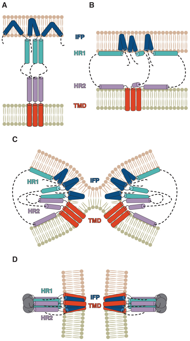
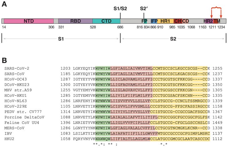
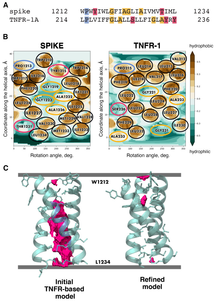
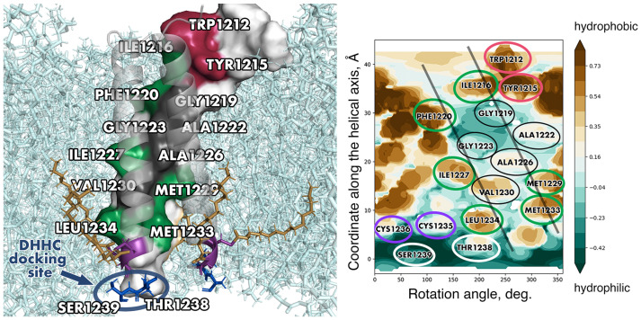
Membrane-spanning portions of proteins' polypeptide chains are commonly known as their transmembrane domains (TMDs). The structural organisation and dynamic behaviour of TMDs from proteins of various families, be that receptors, ion channels, enzymes etc., have been under scrutiny on the part of the scientific community for the last few decades. The reason for such attention is that, apart from their obvious role as an "anchor" in ensuring the correct orientation of the protein's extra-membrane domains (in most cases functionally important), TMDs often actively and directly contribute to the operation of "the protein machine". They are capable of transmitting signals across the membrane, interacting with adjacent TMDs and membrane-proximal domains, as well as with various ligands, etc. Structural data on TMD arrangement are still fragmentary at best due to their complex molecular organisation as, most commonly, dynamic oligomers, as well as due to the challenges related to experimental studies thereof. Inter alia, this is especially true for viral fusion proteins, which have been the focus of numerous studies for quite some time, but have provoked unprecedented interest in view of the SARS-CoV-2 pandemic. However, despite numerous structure-centred studies of the spike (S) protein effectuating target cell entry in coronaviruses, structural data on the TMD as part of the entire spike protein are still incomplete, whereas this segment is known to be crucial to the spike's fusogenic activity. Therefore, in attempting to bring together currently available data on the structure and dynamics of spike proteins' TMDs, the present review aims to tackle a highly pertinent task and contribute to a better understanding of the molecular mechanisms underlying virus-mediated fusion, also offering a rationale for the design of novel efficacious methods for the treatment of infectious diseases caused by SARS-CoV-2 and related viruses.

Keywords: helix–helix interface; membrane protein structure; molecular modelling; protein–protein interactions in membrane; transmembrane helical trimer; viral fusion protein; viral protein acylation.


Conflict of interest statement

The authors declare no conflict of interest.

Figures

Figure 1
Figure 1
Putative coronavirus spike-mediated fusion mechanism. After the dissociation of the S1 subunit, the internal fusion peptide (IFP) is believed to penetrate the target membrane (A). Meanwhile, HR1 and HR2, due to their amphipathic nature, bind to the target membrane and viral envelope, respectively (B). As refolding takes place, HR1 and HR2 form hairpin-like structures with each other, resulting in the membranes approaching each other and merging (C,D), while the protein transitions to the post-fusion state. (Adapted from [12] taking into account the recent results obtained by Shi et al. [10]).
Figure 2
Figure 2
Coronavirus spike proteins and their TMD sequences. (A) Spike protein organisation as exemplified by the spike of SARS-CoV-2. The S1 subunit mainly responsible for target cell recognition comprises the N-terminal (NTD), receptor-binding (RBD) and C-terminal (CTD) domains. Downstream of the S1/S2 cleavage site is the S2 subunit effectuating mostly mechanistic membrane fusion; S2 is additionally cleaved at the S2’ site, immediately followed by the fusion peptide (FP), apart from which S2 includes the internal fusion peptide (IFP), HR1, central helix (CH), connector (CD), HR2 and transmembrane (TM) domains. The region of interest within the scope of our review is located between two red arrows. (Adapted from [19]). (B) Sequence alignment of the fragment from different coronaviruses indicated by arrows in panel (A). The aromatic-rich region, the hydrophobic region and the cysteine-rich region are highlighted in green, red and yellow, respectively. Identical, conserved and semi-conserved residues are designated with the asterisk (*), colon (:) and dot (.) symbols, respectively.
Figure 3
Figure 3
Template-based modelling as applied to the spike TMD. (A) TMD sequence alignment for the spike TMD and one of the candidate templates, tumour growth factor receptor 1A (TNFR-1A). Hydrophobic residues (as well as the charged R) are unhighlighted, while proline, small and polar residues are highlighted in blue, yellow and pink, respectively. (B) Molecular hydrophobicity potential (MHP) distribution maps for an ideal helix corresponding to S-protein residues 1212–1234 and the TMD monomers of candidate template TNFR-1. Cylindrical projection of the surface MHP distribution is used. Axis values correspond to the rotation angle around the helical axis and the distance along the latter, respectively. An MHP scale (in logP octanol-1/water units) is presented on the right. The maps are coloured in accordance with the MHP values [57], from teal (hydrophilic areas) to brown (hydrophobic ones). Projections of proline, small, polar and hydrophobic residues are encircled in blue, yellow, pink and black, respectively. (C) Free volume in the trimer lumen in the initial state built via template-based modelling and in the final trimer refined in the course of MD simulations. Protein chains are partially transparent and are shown in cartoon representation; residues within each chain facing either of the remaining two chains are shown in stick representation. Free volume is rendered as solid pink blocks; membrane boundaries are shown as grey lines.
Figure 4
Figure 4
A uniquely stable model of spike TMD built by Aliper et al. [20]. (Left) The model in 3D. One of the helices is shown in surface representation; the other two helices are shown in cartoon representation. Identical, conserved and non-conserved residues on the helix/helix interface are coloured dark red, green and dark grey, respectively. Palmitoylation sites, palmitoyl tails and lipids are shown in stick representation and coloured purple, dark golden and cyan, respectively. Residues constituting the hypothesised docking site for the enzyme conducting palmitoylation (DHHC) are coloured dark blue, encircled in dark blue and labelled accordingly. (Right) Helix/helix interfaces in one of the chains visualised on a surface MHP map. Identical positions are encircled in red, conserved and semi-conserved residues are encircled in green, and non-conserved residues present on the helix/helix interface are encircled in black. Cys residues and residues constituting the putative docking site for DHHC are encirced in purple and white, respectively. Projections of the other two helices are shown as semi-transparent black lines. For other details, see legend to Figure 3.
Figure 5
Figure 5
The interface between the internal fusion peptide (IFP) and TMD of the spike in the post-fusion state (structure PDB ID 8FDW [10]). (A) Molecular hydrophobicity potential (MHP) distribution maps for the TMD and IFP. See legend to Figure 3 for further detail. Projections of residues on the TMD/IFP interface are encircled in gold. A residue was considered to be on the interface if the area of its solvent-accessible surface went down by at least 25 Å2 compared with the monomeric state (for Gly residues, this difference had to be at least 10 Å2). (B) A 3D structure of the membrane-buried portion of the spike. The TMD and IFP are coloured red and dark blue, while the rest of the protein is coloured beige. The protein is shown in cartoon representation and is semi-transparent apart from one IFP and one TMD interacting with each other, in which residues on the interface are shown in stick representation.
Figure 5
Figure 5
The interface between the internal fusion peptide (IFP) and TMD of the spike in the post-fusion state (structure PDB ID 8FDW [10]). (A) Molecular hydrophobicity potential (MHP) distribution maps for the TMD and IFP. See legend to Figure 3 for further detail. Projections of residues on the TMD/IFP interface are encircled in gold. A residue was considered to be on the interface if the area of its solvent-accessible surface went down by at least 25 Å2 compared with the monomeric state (for Gly residues, this difference had to be at least 10 Å2). (B) A 3D structure of the membrane-buried portion of the spike. The TMD and IFP are coloured red and dark blue, while the rest of the protein is coloured beige. The protein is shown in cartoon representation and is semi-transparent apart from one IFP and one TMD interacting with each other, in which residues on the interface are shown in stick representation.

Similar articles

Cited by

References

    1. Wrobel A.G. Mechanism and evolution of human ACE2 binding by SARS-CoV-2 spike. Curr. Opin. Struct Biol. 2023;81:102619. doi: 10.1016/j.sbi.2023.102619. - DOI - PMC - PubMed
    1. Qi H., Liu B., Wang X., Zhang L. The humoral response and antibodies against SARS-CoV-2 infection. Nat. Immunol. 2022;23:1008–1020. doi: 10.1038/s41590-022-01248-5. - DOI - PubMed
    1. Kordyukova L.V., Shanko A.V. COVID-19: Myths and Reality. Biochemistry. 2021;86:800–817. doi: 10.1134/S0006297921070026. - DOI - PMC - PubMed
    1. Jackson C.B., Farzan M., Chen B., Choe H. Mechanisms of SARS-CoV-2 entry into cells. Nat. Rev. Mol. Cell Biol. 2022;23:3–20. doi: 10.1038/s41580-021-00418-x. - DOI - PMC - PubMed
    1. Basso L.G.M., Zeraik A.E., Felizatti A.P., Costa-Filho A.J. Membranotropic and biological activities of the membrane fusion peptides from SARS-CoV spike glycoprotein: The importance of the complete internal fusion peptide domain. Biochim. Biophys. Acta Biomembr. 2021;1863:183697. doi: 10.1016/j.bbamem.2021.183697. - DOI - PMC - PubMed

LinkOut - more resources