. 2023 Nov 16;24(22):16423.
doi: 10.3390/ijms242216423.

Self-Entrapment of Antimicrobial Peptides in Silica Particles for Stable and Effective Antimicrobial Peptide Delivery System

Affiliations

Self-Entrapment of Antimicrobial Peptides in Silica Particles for Stable and Effective Antimicrobial Peptide Delivery System

Mi-Ran Ki et al. Int J Mol Sci. .

Abstract

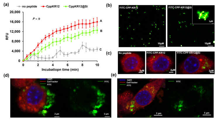
Antimicrobial peptides (AMPs) have emerged as a promising solution to tackle bacterial infections and combat antibiotic resistance. However, their vulnerability to protease degradation and toxicity towards mammalian cells has hindered their clinical application. To overcome these challenges, our study aims to develop a method to enhance the stability and safety of AMPs applicable to effective drug-device combination products. The KR12 antimicrobial peptide was chosen, and in order to further enhance its delivery and efficacy the human immunodeficiency virus TAT protein-derived cell-penetrating peptide (CPP) was fused to form CPP-KR12. A new product, CPP-KR12@Si, was developed by forming silica particles with self-entrapped CPP-KR12 peptide using biomimetic silica precipitability because of its cationic nature. Peptide delivery from CPP-KR12@Si to bacteria and cells was observed at a slightly delivered rate, with improved stability against trypsin treatment and a reduction in cytotoxicity compared to CPP-KR12. Finally, the antimicrobial potential of the CPP-KR12@Si/bone graft substitute (BGS) combination product was demonstrated. CPP-KR12 is coated in the form of submicron-sized particles on the surface of the BGS. Self-entrapped AMP in silica nanoparticles is a safe and effective AMP delivery method that will be useful for developing a drug-device combination product for tissue regeneration.

Keywords: antimicrobial peptide; biomimetic silica deposition; cell penetrating peptide; drug delivery; drug device combination; silica forming peptide.


Conflict of interest statement

The authors declare no conflict of interest.

Figures

Scheme 1
Scheme 1
Self-entrapment of AMPs in silica matrix through CPP-KR12-mediated silica deposition (a) and antimicrobial peptide-device combination product (b).
Figure 1
Figure 1
(a) SYTOXTM Green uptake assay analyzing membrane permeability in the indicated bacterial cells over KR12, CPP, and CPP-KR12. Relative fluorescence unit (RFU) value represents fluorescence ratio relative to that measured in the absence of peptide in each strain after 10 min exposure of indicated peptide (30 μM). Values are presented as the mean ± SE (N = 3). * p < 0.05 vs. KR12 in each strain. (b) Scanning electron microscopy (SEM) image of E. coli treated with indicated AMP. The image is a 10,000× magnification, and the scale bar represents 1 µm. White arrowheads indicate fragmented cells.
Figure 2
Figure 2
Gel retardation assay of peptide–DNA complexes was performed using peptides of varying concentrations. The peptides used for complexation with DNA were (a) KR12, (b) CPP, and (c) CPP-KR12. The concentration of peptide used for complexation with DNA is expressed in micromolar units.
Figure 3
Figure 3
Anti-inflammatory response of AMPs in LPS-stimulated RAW264.7 cells. The mRNA expression levels of inflammatory cytokines, namely TNF-α, IL-1β, and IL-6, were measured by qRT-PCR. The Glyceraldehyde-3-Phosphate-Dehydrogenase (GAPDH) gene was used for normalization in gene expression. The gene levels in each treated cell were calculated relative to those in cells exposed to LPS without AMP. IL-6 was not detected in the control without both LPS and AMP; therefore, it was indicated as nd (not detected). Values are presented as the mean ± SE (N = 3). *** p < 0.001, **** p < 0.0001.
Figure 4
Figure 4
Shape and properties of silica particles formed by each AMP. SEM image at 10,000× magnification (a) and size distribution (b) of silica particles formed by each AMP. The dispersion of particle size was obtained by measuring the diameter of 50 particles from SEM images. The middle line on the figure represents the average sizes for the corresponding particles and the upper and lower lines represent standard deviation. ****p < 0.0001. (c) Zeta potential of each silica particle dispersed in PBS. The zeta potential value is the mean and standard deviation of the three measurements. ***p < 0.001, ****p < 0.0001 vs. Si, #### p < 0.0001 vs. KR12@Si.
Figure 5
Figure 5
Fluorometric determination of the relative sensitivity of suspensions of E. coli to free CPP-KR12 and CPP-KR12@Si with SYTOXTM Green stain. E. coli (108 CFU/mL) was incubated with 5μM of SYTOXTM Green at room temperature for 20 min in 1× PBS. The fluorescence emission spectrum of each 490 nm-excited 100-fold diluted E. coli suspension in the absence or presence of 30 μM of CPP-KR12 or CPP-KR12@Si was acquired for 10 min (a). At the 0.05 level, the mean values for the groups treated with no peptide, CPP-KR12, and CPP-KR12@Si are significantly different from each other (two-way analysis of variance (ANOVA), coupled with Bonferroni post hoc test for multiple comparison). Means that do not share a letter are significantly different. Delivery of FITC-CPP-KR12 to E. coli (b) and to Raw264.7 cells (c). Silica particles were labeled by FITC-conjugated corresponding peptides. Cytosol and nuclei of Raw264.7 were stained by CellTracker Red and 4’,6-diamidino-2-phenylindole (DAPI), respectively. Raw264.7 cells exposed to FITC-CPP-KR12@Si for 2 h (d) and 24 h (e). In (d,e), the left panel displays the merged fluorescence of DAPI, CellTracker, and FITC, which helps identify nuclei, cytosol, and particles. In contrast, the right panel only shows FITC fluorescence, emphasizing the distribution of particles.
Figure 6
Figure 6
Comparison of residual antibacterial activity of AMP and AMP entrapped in silica after trypsin treatment and SEM image of S. aureus treated with CPP-KR12@Si. (a) The residual antibacterial activity was calculated by taking the difference in optical density at 600 nm between the negative control and the sample containing the trypsinized AMP. It was then divided by the optical density of the negative control and multiplied by 100. Each bacterial solution grown in a medium without AMP is used as a corresponding negative control. Values are presented as the mean ± SE (N = 3). * p < 0.05, ** p < 0.01, or *** p < 0.001 vs. corresponding free form of AMP in each strain. (b) SEM image of S. aureus treated with CPP-KR12@Si. The image is a 10,000× magnification, the inside image is a 20,000× magnification, and the scale bar represents 1 µm.
Figure 7
Figure 7
The hemolytic activity and cytotoxicity of AMPs. (a) Hemolysis (%) is the difference in optical density at 570 nm of sheep red blood cells (RBCs) between samples containing the indicated AMP and the negative control as a percentage of the difference in optical density between the positive and negative controls. RBCs present in PBS alone serve as a negative control, whereas RBCs treated with 1% Triton X-100 in PBS serve as a positive control. Values are presented as the mean ± SE (N = 3). ** p < 0.01, *** p < 0.001, or **** p < 0.0001 vs. corresponding free form of AMP in each concentration. (b) The cytotoxicity of AMP was measured 24 h after the addition of each indicated concentration of AMP to Raw264.7 cells grown overnight. Cell survival is expressed as a percentage of the negative control grown without AMP. Values are presented as the mean ± SE (N = 3). *** p < 0.001 or **** p < 0.0001 vs. corresponding free form of AMP in each concentration.
Figure 8
Figure 8
AMP-device combination and its antimicrobial activity. (a) X-ray photoelectron spectroscopy of the as-prepared carriers. Atomic percentages of main elements observed in carriers. The atomic percentages of the constituent were calculated as the ratio of each atom to the sum of the main compounds, except carbon. (b) SEM images of AMP-device combination. The image is a 10,000× magnification. Scale bar: 1 μm. (c) Fluorescence microscope images of E. coli stained with BacLight live/dead kit, which displays green for the live bacteria and red for dead bacteria in medium solutions with the indicated device. Scale bar: 20 μm.

Similar articles

Cited by

References

    1. Uçkay I., Hoffmeyer P., Lew D., Pittet D. Prevention of surgical site infections in orthopaedic surgery and bone trauma: State-of-the-art update. J. Hosp. Infect. 2013;84:5–12. doi: 10.1016/j.jhin.2012.12.014. - DOI - PubMed
    1. Bayramov D.F., Neff J.A. Beyond conventional antibiotics—New directions for combination products to combat biofilm. Adv. Drug Deliv. Rev. 2017;112:48–60. doi: 10.1016/j.addr.2016.07.010. - DOI - PMC - PubMed
    1. Schierholz J.M., Beuth J. Implant infections: A haven for opportunistic bacteria. J. Hosp. Infect. 2001;49:87–93. doi: 10.1053/jhin.2001.1052. - DOI - PubMed
    1. Dostert M., Trimble M.J., Hancock R.E.W. Antibiofilm peptides: Overcoming biofilm-related treatment failure. RSC Adv. 2021;11:2718–2728. doi: 10.1039/D0RA09739J. - DOI - PMC - PubMed
    1. Carpa R., Farkas A., Dobrota C., Butiuc-Keul A. Double-Network Chitosan-Based Hydrogels with Improved Mechanical, Conductive, Antimicrobial, and Antibiofouling Properties. Gels. 2023;9:278. doi: 10.3390/gels9040278. - DOI - PMC - PubMed

LinkOut - more resources