. 2023 Nov 17;24(22):16466.
doi: 10.3390/ijms242216466.

RAR-Dependent and RAR-Independent RXR Signaling in Stem-like Glioma Cells

Affiliations

RAR-Dependent and RAR-Independent RXR Signaling in Stem-like Glioma Cells

Amanda Dabrock et al. Int J Mol Sci. .

Abstract

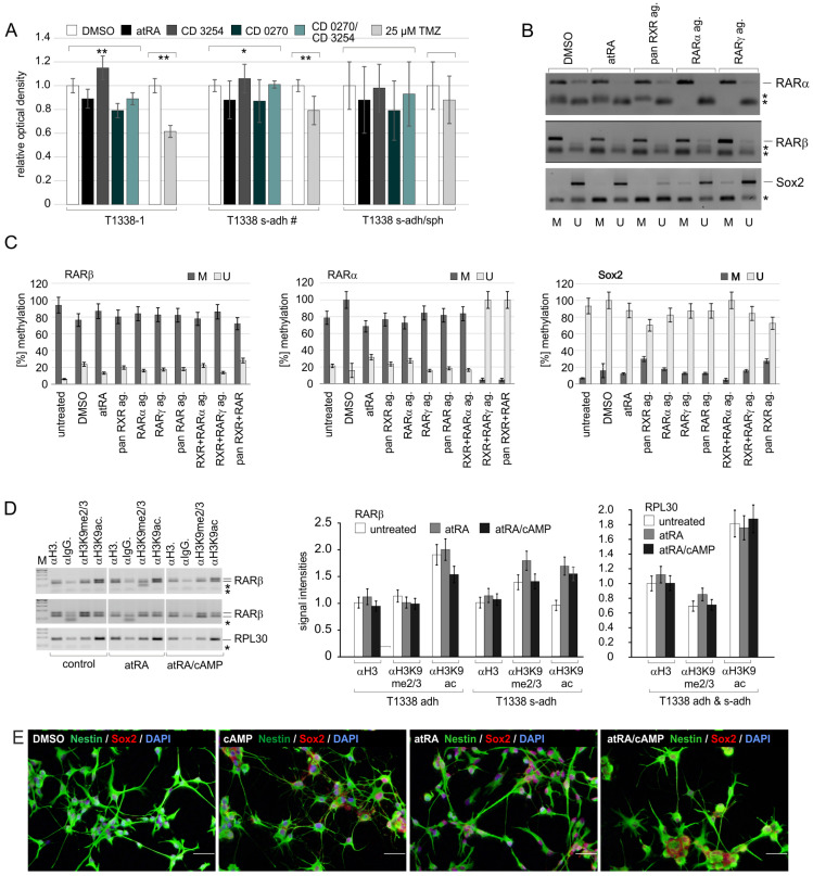
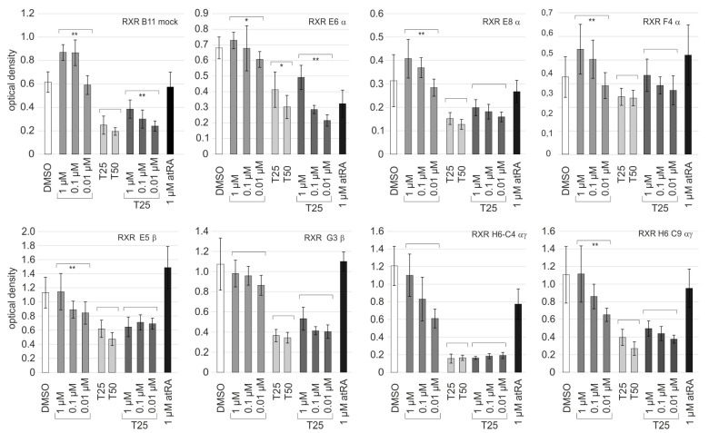
Retinoic acid (RA) exerts pleiotropic effects during neural development and regulates homeostasis in the adult human brain. The RA signal may be transduced through RXR (retinoid-X receptor)-non-permissive RA receptor/RXR heterodimers or through RXR-permissive RXR heterodimers. The significance of RA signaling in malignant brain tumors such as glioblastoma multiforme (GBM) and gliosarcoma (GS) is poorly understood. In particular, the impact RA has on the proliferation, survival, differentiation, or metabolism of GBM- or GS-derived cells with features of stem cells (SLGCs) remains elusive. In the present manuscript, six GBM- and two GS-derived SLGC lines were analyzed for their responsiveness to RAR- and RXR-selective agonists. Inhibition of proliferation and initiation of differentiation were achieved with a RAR-selective pan-agonist in a subgroup of SLGC lines, whereas RXR-selective pan-agonists (rexinoids) supported proliferation in most SLGC lines. To decipher the RAR-dependent and RAR-independent effects of RXR, the genes encoding the RAR or RXR isotypes were functionally inactivated by CRISPR/Cas9-mediated editing in an IDH1-/p53-positive SLGC line with good responsiveness to RA. Stemness, differentiation capacity, and growth behavior were preserved after editing. Taken together, this manuscript provides evidence about the positive impact of RAR-independent RXR signaling on proliferation, survival, and tumor metabolism in SLGCs.

Keywords: CRISPR/Cas9 editing; malignant glioma; retinoic acid receptors; retinoid-X receptor; synthetic retinoids.


Conflict of interest statement

We declare that we have no potential conflict of interest.

Figures

Figure 1
Figure 1
Characteristics of SLGC lines and sensitivity to treatment with rexinoids. (A) Phenotypic characterization of SLGC lines: immunocytochemistry analysis (ICC) using the antibody combination mouse-anti-Sox2/rabbit-anti-CD133. Examples of ICCs using a rabbit-anti-Sox2 antibody are shown in the supplementary figures (e.g., Figure S1A–C). Bars, 50 µm. (B) Growth behavior of distinct T1338 cultures. Sph, spherical growth; s-adh, adherent aggregates; adh, adherent growth. Microphotographs were taken by a life-imaging system. Bars, 200 µm. (C) BrdU ELISA on day d5 using 1 µM atRA or increasing concentrations of the RXR pan-agonist CD3254 (0.01, 10−g M; 0.1, 10−M M; 1, 10−M M). Statistical comparisons to the DMSO control are shown (*, p < 0.05; **, p < 0.001). (D) Variation of BrdU incorporation in the presence of DMSO, 1 µM atRA, 1 µM of the rexinoids CD3254 or Bexarotene, or 1 µM of the RAR pan-agonist CD0270. The values were determined using 15 biological replicates of T1338 cells and included eight independent values per measurement. For the sake of clarity, only the statistical comparisons to the DMSO control are shown (**, p < 0.001). (E) Relative abundance of type I, II, and III cells in the SLGC lines analyzed in the present study. The graphs summarize the data from the ICCs of the biological replicates used for the experiments shown in (C,D). Brackets indicate the statistical comparisons made (**, p < 0.001). Sox2, SRY (sex determining region Y)-box 2 (marker for type I, II, and III cells); CD133, Promin-1 (marker for type II cells).
Figure 2
Figure 2
Characteristics of T1338 subclones. (A) Responsiveness of T1338 subclones to retinoids and TMZ (temozolomide). BrdU incorporation in the presence of DMSO, 1 µM atRA, or 1 µM CD3254, the RAR pan-agonist CD0270 or CD0270/CD3254. The values were determined using three biological replicates and included eight independent values per replicate. Statistical comparisons are indicated by brackets (*, p < 0.05; **, p < 0.001). s-adh #, T1338-2 and -3; s-adh/sph, T1338-9. (B,C) Methylation status of the RARβ, RARα, or Sox2 promoter and impact of ligands. M, methylated; U, unmethylated. The agarose gels (panel B) depict one example of four biological replicates of T1338-1; the symbol * indicates the position of oligo-nucleotides and/or unspecific PCR products. The graphs (panel C) summarize the data from these replicates. Mean values and standard deviations are shown. (D) Chromatin-IP indicating the posttranslational modifications of histone H3 in the RARβ gene promoter: H3K9ac, active mark; H3K9me2/3, repressive mark; ac, acetylated; me2/3, di-/tri-methylated. The agarose gels (left panel) depict the results of two independent biological replicates; the symbol * indicates the position of oligo-nucleotides and/or unspecific PCR products. adh, T1338-6; s-adh, T1338-1 and -2. The bars in the graphs summarize the data from three technical replicates. The IPs with the IgG antibody served as negative controls. (E) Analysis of the differentiation capacity of the biological replicate of T1338-1 used as the mother culture for the editing approaches. Immunocytochemistry analysis using antibodies against Nestin and Sox2 (SRY (sex determining region Y)-box 2) and nuclear counterstain with DAPI (4’,6-Diamidino-2-phenylindol). Bars, 50 µm.
Figure 3
Figure 3
Impact of CRISPR/Cas9-mediated editing of the RAR genes. (A) Left: schematic drawing indicating the two zinc fingers of the RAR DBDs and the P box (-CEGCK-). The black arrow indicates the position of editing. Right: amino acid sequences of the first zinc finger (underlined) of RARα, β, and γ; zinc complexing cysteine residues are shown in blue. In general, the editing process eliminated the amino acids C-terminally of the residues indicated in red (for details, see Figures S4, S5 and S6A–C). WT, wildtype; ed*, edited clones. (B) Immunocytochemistry analyses of edited RAR clones using antibodies against Sox2 and CD133. DAPI nuclear counterstain was omitted from the panel RAR E5α. Bars, 50 µm. (C) Similar analysis as in (B) investigating the differentiation capacity of the clones indicated. Bars, 50 µm. (D) Quantification of the presence of type I, II, and III cells in proliferating clones and after induction of differentiation with a RAR pan-agonist. Data from mock controls and RAR clones with the same type of editing (ed.) were combined. Mean values are indicated by a black line. (E) Growth curves of three edited clones and one mock-edited clone in the presence of 1 µM atRA or the carrier DMSO. Cell numbers were determined on days d2, d5, d8, and d12. The mean values of four independent assays are shown; whiskers indicate standard deviations; statistical comparisons are indicated by brackets (**, p < 0.001). For the sake of clarity, only statistical comparisons between the treated and untreated samples from the same day are shown. β* indicates monoallelic editing of the RARB gene.
Figure 4
Figure 4
Impact of CRISPR/Cas9-mediated editing of the RXR genes. (A) Left: schematic drawing indicating the two zinc fingers of the RXR DBDs and the P box (-CEGCK-). The black arrow indicates the position of editing. Right: amino acid sequences of the first zinc finger (underlined) of RXRα, β, and γ; zinc complexing cysteine residues are shown in blue. In general, the editing process eliminated the amino acids C-terminally of the residues indicated in red (for details, see Figures S7 and S8A–C). (B) Immunocytochemistry analyses of the stemness state and DAPI nuclear counterstain. Bars, 50 µm. (C) Quantification of the presence of type I, II, and III cells in edited clones (ed.) and impact of the induction of differentiation (diff). Mock controls and RXR clones with the same type of knockout are summarized in one graph each. Data from 2–3 biological replicates are combined. Mean values are indicated by a black line. (D) A similar experiment as in (B) investigating the capacity for differentiation. Bars, 50 µm. (E) Growth curves of seven edited and two mock clones. Cell numbers were determined on days d2, d5, d8, and d12. The mean values of five independent replicates are shown; whiskers indicate standard deviations. Statistical comparisons are indicated by brackets (*, p < 0.05; **, p < 0.001). For the sake of clarity, only statistical comparisons between the samples from the same day are shown.
Figure 4
Figure 4
Impact of CRISPR/Cas9-mediated editing of the RXR genes. (A) Left: schematic drawing indicating the two zinc fingers of the RXR DBDs and the P box (-CEGCK-). The black arrow indicates the position of editing. Right: amino acid sequences of the first zinc finger (underlined) of RXRα, β, and γ; zinc complexing cysteine residues are shown in blue. In general, the editing process eliminated the amino acids C-terminally of the residues indicated in red (for details, see Figures S7 and S8A–C). (B) Immunocytochemistry analyses of the stemness state and DAPI nuclear counterstain. Bars, 50 µm. (C) Quantification of the presence of type I, II, and III cells in edited clones (ed.) and impact of the induction of differentiation (diff). Mock controls and RXR clones with the same type of knockout are summarized in one graph each. Data from 2–3 biological replicates are combined. Mean values are indicated by a black line. (D) A similar experiment as in (B) investigating the capacity for differentiation. Bars, 50 µm. (E) Growth curves of seven edited and two mock clones. Cell numbers were determined on days d2, d5, d8, and d12. The mean values of five independent replicates are shown; whiskers indicate standard deviations. Statistical comparisons are indicated by brackets (*, p < 0.05; **, p < 0.001). For the sake of clarity, only statistical comparisons between the samples from the same day are shown.
Figure 5
Figure 5
Characteristics of clones with edited RXR genes. (A) BrdU-ELISA on day d5: comparison of eight edited clones and one mock clone (B11) and impact of the treatment with 1µM of CD3254. The mean of eight independent values and the standard deviations are shown. The brackets indicate statistical comparisons (*, p < 0.05; **, p < 0.001). (B) Immunocytochemistry analyses using the antibody combination GFAP/Tau and DAPI nuclear counterstain. Expression after treatment with 1 µM atRA/1 mM cAMP and in the corresponding controls. Bars, 50 µm. cAMP (cyclic adenosine monophosphate).
Figure 6
Figure 6
Impact of RXR editing on the expression of metabolic enzymes. Expression of the pyruvate kinase isoform PKM2 and the lactate dehydrogenase isoform A (LDHA) in RXR clones. Left: an example of Western blot analysis; the loading control Actin is shown below. Middle and right: The graphs depict the quantification of the relative expression of the PKM2 and the LDHA, respectively. Significantly distinct expression levels are indicated by ** (p < 0.001).
Figure 7
Figure 7
Impact of treatment with TMZ in the presence of bexarotene. BrdU-ELISA on day d5: Comparison of the proliferation of seven edited clones and one mock-edited clone (B11) after treatment with increasing concentrations of bexarotene (1 µM, 0.1 µM, and 0.01 µM) in the absence (light gray bars) and presence (dark gray bars) of 25 µM TMZ (T25). Treatment with 1 µM atRA (black bars), TMZ (T25, 25 µM; T50, 50 µM), or the carrier DMSO (white bars) served as controls. The mean of eight independent values and the standard deviations are shown. For the sake of clarity, only statistical comparisons between the samples from the same treatment groups are shown (*, p < 0.05; **, p < 0.001).
Figure 8
Figure 8
Impact of the treatment with TMZ and Bexarotene on cell numbers and stemness. (A) Relative expression of Sox2 (or p21CIP1 or MGMT) in clones with edited RXRα (α−/−; E6α, E8α, E10α, F4α), RXRβ (β−/−; E5β, G3β, E9β), RXRγ (γ−/−), as well as clones with no editing (mock) and a double knockout (RXRαγ−/−; H6, H6C9, H6C4); the impacts of 25 µM TMZ and 1 µM bexarotene are shown. A representative example of a Western blot is depicted in the middle. Expression of p21 (p21CIP1) was low and varied between replicates of the same clones and clones with the same type of knockout. Brackets indicate the statistical comparisons performed (**, p < 0.001). (B,C) Quantification of the cell numbers revealed by immunocytochemistry analyses using the antibody combinations CD133/Sox2 (A) or Nestin/γH2AX (B). The mean values and standard deviations are shown. Brackets indicate the statistical comparisons performed (**, p < 0.001). In (B), the relative amounts of Sox2-negative cells are indicated in blue, and in (C), the relative proportion of γH2AX-positive cells is indicated in green. A robust quantification of type I, II, and III cells was not possible because of staining artifacts in TMZ-treated cell batches.

References

    1. Stupp R., Hegi M.E., Gilbert M.R., Chakravarti A. Chemoradiotherapy in Malignant Glioma: Standard of Care and Future Directions. J. Clin. Oncol. 2007;25:4127–4136. doi: 10.1200/JCO.2007.11.8554. - DOI - PubMed
    1. The Cancer Genome Atlas Research Network Comprehensive genomic characterization defines human glioblastoma genes and core pathways. Nature. 2008;455:1061–1068. doi: 10.1038/nature07385. - DOI - PMC - PubMed
    1. Nakada M., Kita D., Watanabe T., Hayashi Y., Teng L., Pyko I.V., Hamada J. Aberrant signaling pathways in glioma. Cancers. 2011;3:3242–3278. doi: 10.3390/cancers3033242. - DOI - PMC - PubMed
    1. Louis D.N., Perry A., Reifenberger G., von Deimling A., Figarella-Branger D., Cavenee W.K., Ohgaki H., Wiestler O.D., Kleihues P., Ellison D.W. The 2016 World Health Organization Classification of Tumors of the Central Nervous System: A summary. Acta Neuropathol. 2016;131:803–820. doi: 10.1007/s00401-016-1545-1. - DOI - PubMed
    1. Beier D., Schulz J.B., Beier C.P. Chemoresistance of glioblastoma cancer stem cells—Much more complex than expected. Mol. Cancer. 2011;10:128. doi: 10.1186/1476-4598-10-128. - DOI - PMC - PubMed