. 2023 Nov 20;24(22):16520.
doi: 10.3390/ijms242216520.

Interaction of TSG101 with the PTAP Motif in Distinct Locations of Gag Determines the Incorporation of HTLV-1 Env into the Retroviral Virion

Affiliations

Interaction of TSG101 with the PTAP Motif in Distinct Locations of Gag Determines the Incorporation of HTLV-1 Env into the Retroviral Virion

Yosuke Maeda et al. Int J Mol Sci. .

Abstract

Human T-cell tropic virus type 1 (HTLV-1) is known to be mainly transmitted by cell-to-cell contact due to the lower infectivity of the cell-free virion. However, the reasons why cell-free HTLV-1 infection is poor remain unknown. In this study, we found that the retrovirus pseudotyped with HTLV-1 viral envelope glycoprotein (Env) was infectious when human immunodeficiency virus type 1 (HIV-1) was used to produce the virus. We found that the incorporation of HTLV-1 Env into virus-like particles (VLPs) was low when HTLV-1 Gag was used to produce VLPs, whereas VLPs produced using HIV-1 Gag efficiently incorporated HTLV-1 Env. The production of VLPs using Gag chimeras between HTLV-1 and HIV-1 Gag and deletion mutants of HIV-1 Gag showed that the p6 domain of HIV-1 Gag was responsible for the efficient incorporation of HTLV-1 Env into the VLPs. Further mutagenic analyses of the p6 domain of HIV-1 Gag revealed that the PTAP motif in the p6 domain of HIV-1 Gag facilitates the incorporation of HTLV-1 Env into VLPs. Since the PTAP motif is known to interact with tumor susceptibility gene 101 (TSG101) during the budding process, we evaluated the effect of TSG101 knockdown on the incorporation of HTLV-1 Env into VLPs. We found that TSG101 knockdown suppressed the incorporation of HTLV-1 Env into VLPs and decreased the infectivity of cell-free HIV-1 pseudotyped with HTLV-1 Env. Our results suggest that the interaction of TSG101 with the PTAP motif of the retroviral L domain is involved not only in the budding process but also in the efficient incorporation of HTLV-1 Env into the cell-free virus.

Keywords: HTLV-1; PTAP; TSG101; infectivity; retroviral Gag.


Conflict of interest statement

The authors declare no conflict of interest.

Figures

Figure 1
Figure 1
Cell-free and cell-to-cell infection by HIV-1 and HTLV-1, and pseudotyped virus infection with different Envs. (A) The same volume of culture supernatants from 293T cells transfected with HTLV-1-inGLuc and HIV-1-inGLuc and their respective packaging vectors and Env expression plasmid were used for the cell-free infection of NP-2/CD4/CCR5/CXCR4 cells. Transfected cells were added to NP-2/CD4/CCR5/CXCR4 cells for cell-to-cell infection. The Gaussia luciferase activity of cell-free and cell-to-cell infection was measured after 48 h of infection. The cell-free virus infectivity was normalized by luciferase activity per 1 ng p24 Ag of HTLV-1 and HIV-1, respectively. The untransfected 293T cells were used as the no-virus control. The column and bar indicate the mean and standard deviation in triplicate experiments, respectively (** p < 0.01, *** p < 0.001 using unpaired t-test). (B) Pseudotyped virus produced from 293T cells transfected with HTLV-1-inGLuc or HIV-1-inGLuc vectors; different Envs were used for the cell-free infection of NP-2/CD4/CCR5/CXCR4 cells. The Gaussia luciferase activities of culture supernatant from infected cells were measured after 48 h infection. The pseudotyped virus infectivity was normalized by luciferase activity per 1 ng p24 Ag of HTLV-1 and HIV-1, respectively. The column and bar indicate the mean and standard deviation in triplicate experiments, respectively (* p < 0.05, ** p < 0.01 using unpaired t-test).
Figure 2
Figure 2
HTLV-1 Env expression level and fusion activity in the absence or presence of retroviral Gag, and efficiency of HLTV-1 Env incorporation into VLPs produced by retroviral Gag. (A) The HTLV-1 Env expression plasmid was transfected into 293T cells with control Venus vector (orange), HTLV-1-Gag-Venus (blue), and HIV-1-Gag-Venus (green). After 24 h of transfection, the cells were recovered and stained with a monoclonal antibody against gp46, LAT-27. Venus-positive cell populations were analyzed by flow cytometry. Red indicates a lack of antibody staining (control). (B) The HTLV-1 Env expression plasmid was transfected into 293T cells with control FLAG vector, HTLV-1-Gag-FLAG, and HIV-1-Gag-FLAG plasmids. After 24 h of transfection, transfected cells were recovered and added to TZM-bl cells. The luciferase activities of mixed cells were measured after 24 h of co-culture. The column and bar indicate the mean and standard deviation in quadruplicate experiments, respectively (*** p < 0.001 using unpaired t-test). (C) Cell lysates and VLPs produced from 293T cells transfected with control vector, HTLV-1-Gag-FLAG, or HIV-1-Gag-FLAG combined with HTLV-1 Env were analyzed by Western blotting using anti-FLAG, LAT-27, and anti-CypA antibodies. The arrowhead shows the ~53-kDa size of the Env protein. The positions of the molecular mass marker (kDa) are indicated on the right.
Figure 3
Figure 3
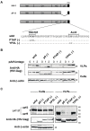
Efficiency of HTLV-1 Env incorporation into VLPs produced in chimeras between HTLV-1 and HIV-1 Gag and mutants in HTLV-1 and HIV-1 Gag. (A) Schematic representation of chimeras between HTLV-1 and HIV-1 Gag and mutants in HTLV-1 and HIV-1 Gag. (B) The cell lysates and VLPs produced from 293T cells transfected with HTLV-1 Env and chimeras between HTLV-1 and HIV-1 Gag or mutants in HTLV-1 and HIV-1 Gag were analyzed by Western blotting using anti-FLAG, LAT-27, and anti-CypA antibodies. The arrowhead shows the ~53-kDa size of the Env protein. The positions of the molecular mass marker (kDa) are indicated on the right.
Figure 4
Figure 4
Efficiency of HTLV-1 Env incorporation into VLPs produced in HIV-1 p6 Gag mutants. (A) Schematic representation of the deletions and mutations in the p6 domain of HIV-1-Gag. Amino acid sequences of the wild-type p6 domain are shown with the PTAP and YPXL motifs. A dash denotes amino acid identity. (B) Cell lysates and VLPs produced from 293T cells transfected with 1 μg of p6 Gag deletion and mutants with different amounts of pAdVantage (0, 1, and 2 μg) were analyzed by Western blotting using anti-HA and anti-β-actin antibodies. (C) Cell lysates and VLPs produced from 293T cells transfected with 1.5 μg of HTLV-1 Env and 3.5 μg of p6 deletion and mutants of HIV-1-Gag-HA were analyzed by Western blotting using anti-HA, LAT-27, and anti-β-actin antibodies. The arrowhead shows the ~53-kDa size of the Env protein. The positions of the molecular mass marker (kDa) are indicated on the right.
Figure 5
Figure 5
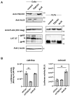
Incorporation of HTLV-1 Env in HIV-1-Gag VLPs and cell-free and cell-to-cell infectivity of HIV-1 Gag pseudotyped with HTLV-1 Env produced in 293T cells with TSG101 or ALIX knockdown. (A) Expression levels of TSG101 and ALIX in 293T cells transduced with shRNA targeting control, TSG101, and ALIX were analyzed by Western blotting using anti-TSG101 and anti-ALIX antibodies. The knockdown 293T cells were transfected with HIV-Gag-FLAG and HLTV-1 Env, and the resulting cell lysates and VLPs were recovered after 48 h of transfection and analyzed by Western blotting using anti-FLAG, LAT-27, and anti-β-actin antibodies. The arrowhead shows the ~53-kDa size of the Env protein. The positions of the molecular mass marker (kDa) are indicated on the right. (B) TSG101 or ALIX knockdown 293T cells were used to produce HIV-1 in GLuc with HIV-1 packaging vectors and the HTLV-1 Env expression plasmid. The same volume of culture supernatant of transfected cells was used for the cell-free infection of NP-2/CD4/CCR5/CXCR4 cells. Transfected cells were added to NP-2/CD4/CCR5/CXCR4 cells for cell-to-cell infection. The Gaussia luciferase activity of the resulting culture supernatant was measured after 48 h of culturing for cell-free and cell-to-cell infection. The cell-free virus infectivity was normalized by luciferase activity per 1 ng p24 Ag of HIV-1. The 293T cells transduced with a control shRNA vector (shCon) were used as a control. The column and bar show the mean and standard deviation in triplicate experiments, respectively (** p < 0.01 using unpaired t-test).

Similar articles

Cited by

References

    1. Gallo R.C. The discovery of the first human retrovirus: HTLV-1 and HTLV-2. Retrovirology. 2005;2:17. doi: 10.1186/1742-4690-2-17. - DOI - PMC - PubMed
    1. Hinuma Y., Nagata K., Hanaoka M., Nakai M., Matsumoto T., Kinoshita K.I., Shirakawa S., Miyoshi I. Adult T-cell leukemia: Antigen in an ATL cell line and detection of antibodies to the antigen in human sera. Proc. Natl. Acad. Sci. USA. 1981;78:6476–6480. doi: 10.1073/pnas.78.10.6476. - DOI - PMC - PubMed
    1. Popovic M., Sarin P.S., Robert-Gurroff M., Kalyanaraman V.S., Mann D., Minowada J., Gallo R.C. Isolation and transmission of human retrovirus (human T-cell leukemia virus) Science. 1983;219:856–859. doi: 10.1126/science.6600519. - DOI - PubMed
    1. Takatsuki K. Discovery of adult T-cell leukemia. Retrovirology. 2005;2:16. doi: 10.1186/1742-4690-2-16. - DOI - PMC - PubMed
    1. Gessain A., Vernant J.C., Maurs L., Barin F., Gout O., Calender A., De Thé G. Antibodies to human T-lymphotropic virus type-I in patients with tropical spastic paraparesis. Lancet. 1985;326:407–410. doi: 10.1016/S0140-6736(85)92734-5. - DOI - PubMed

MeSH terms

LinkOut - more resources