. 2023 Nov 20;24(22):16537.
doi: 10.3390/ijms242216537.

Leucine-Rich Alpha-2 Glycoprotein 1 Accumulates in Complicated Atherosclerosis and Promotes Calcification

Affiliations

Leucine-Rich Alpha-2 Glycoprotein 1 Accumulates in Complicated Atherosclerosis and Promotes Calcification

Lucile Grzesiak et al. Int J Mol Sci. .

Abstract

Atherosclerosis is the primary cause of cardiovascular disease. The development of plaque complications, such as calcification and neo-angiogenesis, strongly impacts plaque stability and is a good predictor of mortality in patients with atherosclerosis. Despite well-known risk factors of plaque complications, such as diabetes mellitus and chronic kidney disease, the mechanisms involved are not fully understood. We and others have identified that the concentration of circulating leucine-rich α-2 glycoprotein 1 (LRG1) was increased in diabetic and chronic kidney disease patients. Using apolipoprotein E knockout mice (ApoE-/-) (fed with Western diet) that developed advanced atherosclerosis and using human carotid endarterectomy, we showed that LRG1 accumulated into an atherosclerotic plaque, preferentially in calcified areas. We then investigated the possible origin of LRG1 and its functions on vascular cells and found that LRG1 expression was specifically enhanced in endothelial cells via inflammatory mediators and not in vascular smooth muscle cells (VSMC). Moreover, we identified that LRG1 was able to induce calcification and SMAD1/5-signaling pathways in VSMC. In conclusion, our results identified for the first time that LRG1 is a direct contributor to vascular calcification and suggest a role of this molecule in the development of plaque complications in patients with atherosclerosis.

Keywords: LRG1; atherosclerosis; calcification; vascular smooth muscle cell.


Conflict of interest statement

The authors declare no conflict of interest.

Figures
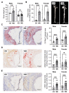
Figure 1
Figure 1
LRG1 in calcified atherosclerotic plaques in a mouse model of advanced atherosclerosis. (A) Body weights in male (n = 9–11/group) and female (n = 18/group) ApoE−/− mice after 27 weeks of Western diet (WD) or normal chow diet (CD). (B) Quantification of the plaque area and representative images of en face aortas for CD and WD-fed male and female mice (n = 9–11/group). (CE) Representative histological images and quantitative analysis in serial sections of aortic sinus from CD and WD-fed male and female mice showing plaque lipid content with Oil Red O coloration (n = 9–11/group) ((C) red staining), plaque calcification with the Alizarin Red staining (n = 16–18/group) ((D) dark red staining) and LRG1 expression (n = 16–18/group) ((E) brown staining). Scoring for Alizarin Red and LRG1 was performed with 3 scores: 0 (no staining), 1 (weak staining), and 2 (strong localized staining). ** p < 0.01, *** p < 0.001, **** p < 0.0001 via Mann–Whitney U test for (A,C,D) and Student’s t-test for (B,E). Normally distributed data are presented as mean ± SEM, and non-normally distributed data are presented as median with lower and upper quartiles.
Figure 2
Figure 2
LRG1 in calcified atherosclerotic plaques in humans. Histological images of human carotid plaque serial sections showing (A) plaque calcification with the Alizarin Red staining (dark red staining) and (B) LRG1 expression (brown staining).
Figure 3
Figure 3
LRG1 expression in endothelial cells and VSMC in response to inflammatory stimuli. Quantification of LRG1-relative mRNA expression in (A,B) human umbilical vein endothelial cells HUVECs, (C,D) murine vascular smooth muscle cells MOVAS, and (E,F) human vascular smooth muscle cells hASMCs stimulated for 24 h and 48 h with 30 ng/mL of TNFα or 4 ng/mL of IL1β. The expression levels of LRG1 mRNA were corrected with Rpl13 expression. N = 3–5 independent experiments/group; AU: Arbitrary Unit; * p < 0.05, ** p < 0.01 by Kruskal–Wallis with Dunn’s multiple comparison test. Data are presented as median with lower and upper quartiles.
Figure 4
Figure 4
Effect of LRG1 in VSMC trans-differentiation and calcification. (A,B) Representative images and quantification of Alizarin Red staining (A) and quantification of calcium deposition (B) in MOVAS treated with control (−Pi) or calcification medium (+Pi) with or without 20 μg/mL LRG1 (+LRG1) for 6 days (n = 15 replicates/group from 5 independent experiments). (C) Quantification of osteoblast-like cells phenotype markers relative mRNA expression Runx2, Spp1, and Ankh in MOVAS treated with control (−Pi) or calcification medium (+Pi) with or without 20 μg/mL LRG1 (+LRG1) for 6 days (n = 4 independent experiments/group). * p < 0.05, *** p < 0.001 by Mann–Whitney U test for (A,C) and Student’s t-test for (B). Normally distributed data are presented as mean ± SEM and non-normally distributed data are presented as median with lower and upper quartiles.
Figure 5
Figure 5
Effect of LRG1 on TGFβ-induced SMAD signaling. (A,B) Quantification and representative capillary Western blots of pSMAD1-5/SMAD1 and (B) pSMAD2-3/SMAD2 in MOVAS treated with 20 μg/mL LRG1 with or without 10 ng/mL TGFβ for 1 h. n = 3–5 independent experiments/group; ns: non significative; ** p < 0.01 via Mann–Whitney U test. Data are presented as median with lower and upper quartiles.

References

    1. Lusis A.J. Atherosclerosis. Nature. 2000;407:233–241. doi: 10.1038/35025203. - DOI - PMC - PubMed
    1. Redgrave J.N., Gallagher P., Lovett J.K., Rothwell P.M. Critical Cap Thickness and Rupture in Symptomatic Carotid Plaques. Stroke. 2008;39:1722–1729. doi: 10.1161/STROKEAHA.107.507988. - DOI - PubMed
    1. Bentzon J.F., Otsuka F., Virmani R., Falk E. Mechanisms of Plaque Formation and Rupture. Circ. Res. 2014;114:1852–1866. doi: 10.1161/CIRCRESAHA.114.302721. - DOI - PubMed
    1. Hutcheson J., Maldonado N., Aikawa E. Small Entities with Large Impact: Microcalcifications and Atherosclerotic Plaque Vulnerability. Curr. Opin. Lipidol. 2014;25:327–332. doi: 10.1097/MOL.0000000000000105. - DOI - PMC - PubMed
    1. Van Rosendael A.R., Narula J., Lin F.Y., van den Hoogen I.J., Gianni U., Al Hussein Alawamlh O., Dunham P.C., Peña J.M., Lee S.-E., Andreini D., et al. Association of High-Density Calcified 1K Plaque with Risk of Acute Coronary Syndrome. JAMA Cardiol. 2020;5:282–290. doi: 10.1001/jamacardio.2019.5315. - DOI - PMC - PubMed