

Collected Thoughts on Mycobacterial Lipoarabinomannan, a Cell Envelope Lipoglycan
- PMID: 38003746
- PMCID: PMC10675199
- DOI: 10.3390/pathogens12111281
Collected Thoughts on Mycobacterial Lipoarabinomannan, a Cell Envelope Lipoglycan
Abstract
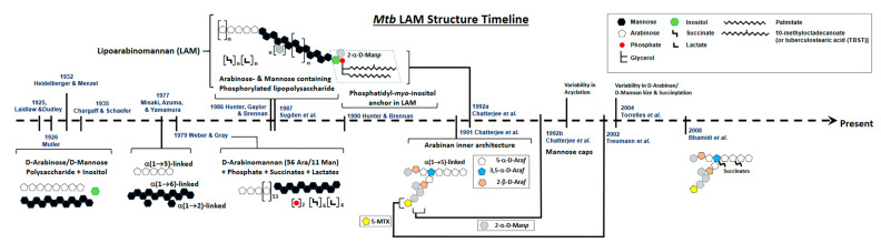
The presence of lipoarabinomannan (LAM) in the Mycobacterium tuberculosis (Mtb) cell envelope was first reported close to 100 years ago. Since then, numerous studies have been dedicated to the isolation, purification, structural definition, and elucidation of the biological properties of Mtb LAM. In this review, we present a brief historical perspective on the discovery of Mtb LAM and the herculean efforts devoted to structurally characterizing the molecule because of its unique structural and biological features. The significance of LAM remains high to this date, mainly due to its distinct immunological properties in conjunction with its role as a biomarker for diagnostic tests due to its identification in urine, and thus can serve as a point-of-care diagnostic test for tuberculosis (TB). In recent decades, LAM has been thoroughly studied and massive amounts of information on this intriguing molecule are now available. In this review, we give the readers a historical perspective and an update on the current knowledge of LAM with information on the inherent carbohydrate composition, which is unique due to the often puzzling sugar residues that are specifically found on LAM. We then guide the readers through the complex and myriad immunological outcomes, which are strictly dependent on LAM's chemical structure. Furthermore, we present issues that remain unresolved and represent the immediate future of LAM research. Addressing the chemistry, functions, and roles of LAM will lead to innovative ways to manipulate the processes that involve this controversial and fascinating biomolecule.
Keywords: Mycobacterium tuberculosis; lipoarabinomannan; lipoglycan; mycobacteria; tuberculosis.
Conflict of interest statement
The authors declare no conflict of interest.
Figures


Similar articles
-
A Journey from Structure to Function of Bacterial Lipopolysaccharides.Chem Rev. 2022 Oct 26;122(20):15767-15821. doi: 10.1021/acs.chemrev.0c01321. Epub 2021 Jul 21. Chem Rev. 2022. PMID: 34286971 Review.
-
Multi-technique-based electrochemical sensing of lipoarabinomannan (LAM) antigen as a biomarker for early-stage tuberculosis diagnosis.Nanotechnology. 2025 Mar 3;36(15). doi: 10.1088/1361-6528/adb7ea. Nanotechnology. 2025. PMID: 39970481
-
Estimation of D-Arabinose by Gas Chromatography/Mass Spectrometry as Surrogate for Mycobacterial Lipoarabinomannan in Human Urine.PLoS One. 2015 Dec 3;10(12):e0144088. doi: 10.1371/journal.pone.0144088. eCollection 2015. PLoS One. 2015. PMID: 26633829 Free PMC article.
-
Lipoarabinomannan regulates septation in Mycobacterium smegmatis.bioRxiv [Preprint]. 2023 Mar 26:2023.03.26.534150. doi: 10.1101/2023.03.26.534150. bioRxiv. 2023. Update in: Nat Commun. 2024 Mar 11;15(1):2191. doi: 10.1038/s41467-024-46565-5. PMID: 36993273 Free PMC article. Updated. Preprint.
-
Value of urine-based lipoarabinomannan (LAM) antigen tests for diagnosing tuberculosis in children: systematic review and meta-analysis.IJID Reg. 2022 Jun 23;4:97-104. doi: 10.1016/j.ijregi.2022.06.004. eCollection 2022 Sep. IJID Reg. 2022. PMID: 35880002 Free PMC article. Review.
Cited by
-
ZnI2-Mediated cis-Glycosylations of Various Constrained Glycosyl Donors: Recent Advances in cis-Selective Glycosylations.Molecules. 2024 Oct 4;29(19):4710. doi: 10.3390/molecules29194710. Molecules. 2024. PMID: 39407638 Free PMC article. Review.
-
Selective Glycan Labeling of Mannose-Containing Glycolipids in Mycobacteria.J Am Chem Soc. 2024 Jan 10;146(1):377-385. doi: 10.1021/jacs.3c09495. Epub 2023 Dec 19. J Am Chem Soc. 2024. PMID: 38112296 Free PMC article.
-
Prospective Analysis of urINe LAM to Eliminate NTM Sputum Screening (PAINLESS) study: Rationale and trial design for testing urine lipoarabinomannan as a marker of NTM lung infection in cystic fibrosis.PLoS One. 2025 Mar 10;20(3):e0309191. doi: 10.1371/journal.pone.0309191. eCollection 2025. PLoS One. 2025. PMID: 40063876 Free PMC article.
-
Long non-coding RNA transcripts in Mycobacterium tuberculosis-host interactions.Noncoding RNA Res. 2024 Dec 15;11:281-293. doi: 10.1016/j.ncrna.2024.12.005. eCollection 2025 Apr. Noncoding RNA Res. 2024. PMID: 39926616 Free PMC article. Review.
-
Prospective Analysis of urINe LAM to Eliminate NTM Sputum Screening (PAINLESS) study: Rationale and trial design for testing urine lipoarabinomannan as a marker of NTM lung infection in cystic fibrosis.medRxiv [Preprint]. 2024 Aug 9:2024.08.08.24311698. doi: 10.1101/2024.08.08.24311698. medRxiv. 2024. Update in: PLoS One. 2025 Mar 10;20(3):e0309191. doi: 10.1371/journal.pone.0309191. PMID: 39148848 Free PMC article. Updated. Preprint.
References
Publication types
Grants and funding
LinkOut - more resources
Full Text Sources
