. 2023 Nov 18;12(11):1367.
doi: 10.3390/pathogens12111367.

Implications of Flagellar Attachment Zone Proteins TcGP72 and TcFLA-1BP in Morphology, Proliferation, and Intracellular Dynamics in Trypanosoma cruzi

Affiliations

Implications of Flagellar Attachment Zone Proteins TcGP72 and TcFLA-1BP in Morphology, Proliferation, and Intracellular Dynamics in Trypanosoma cruzi

Normanda Souza-Melo et al. Pathogens. .

Abstract

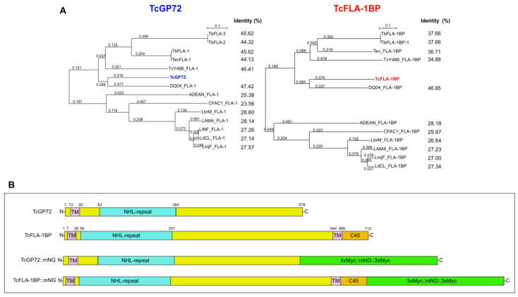
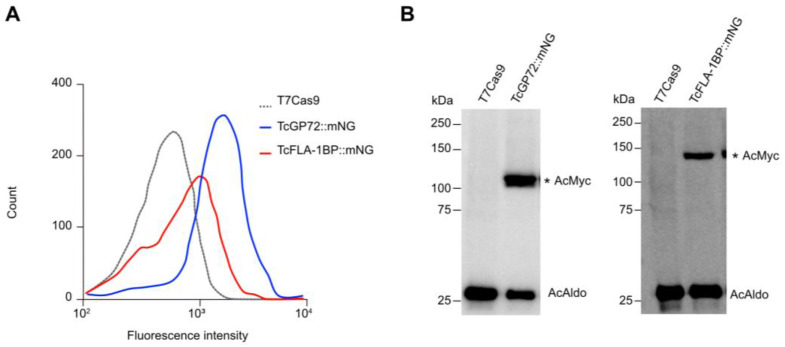
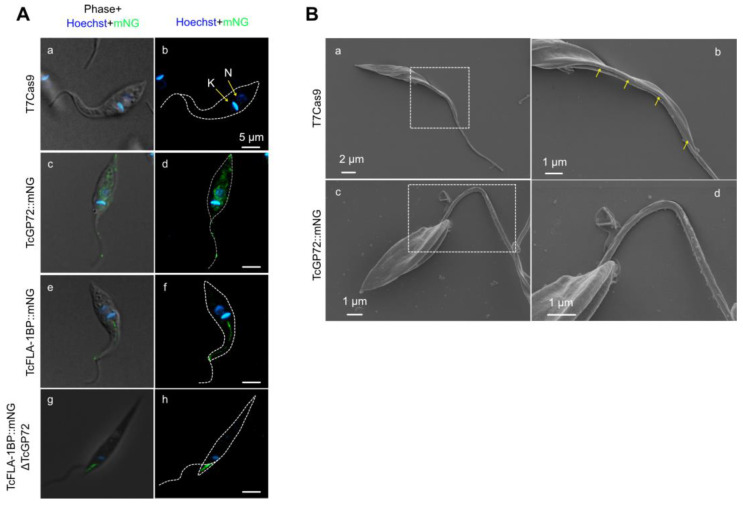
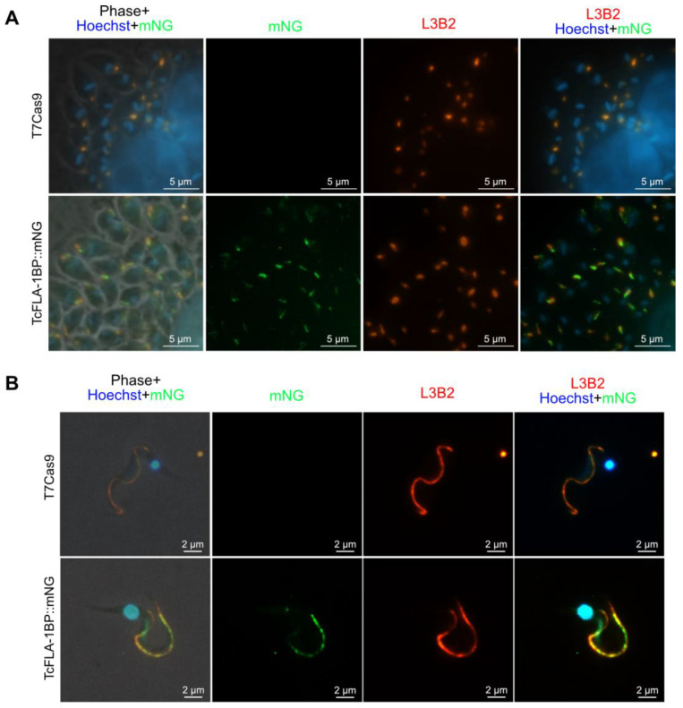
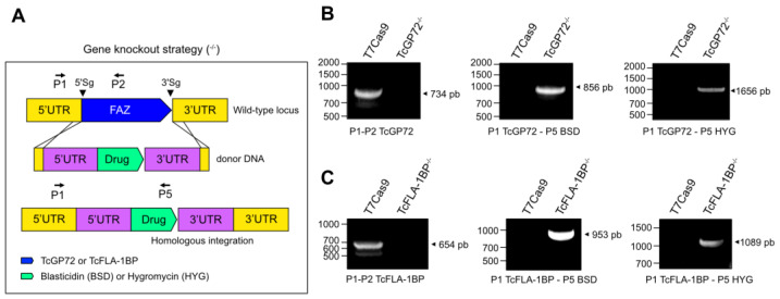
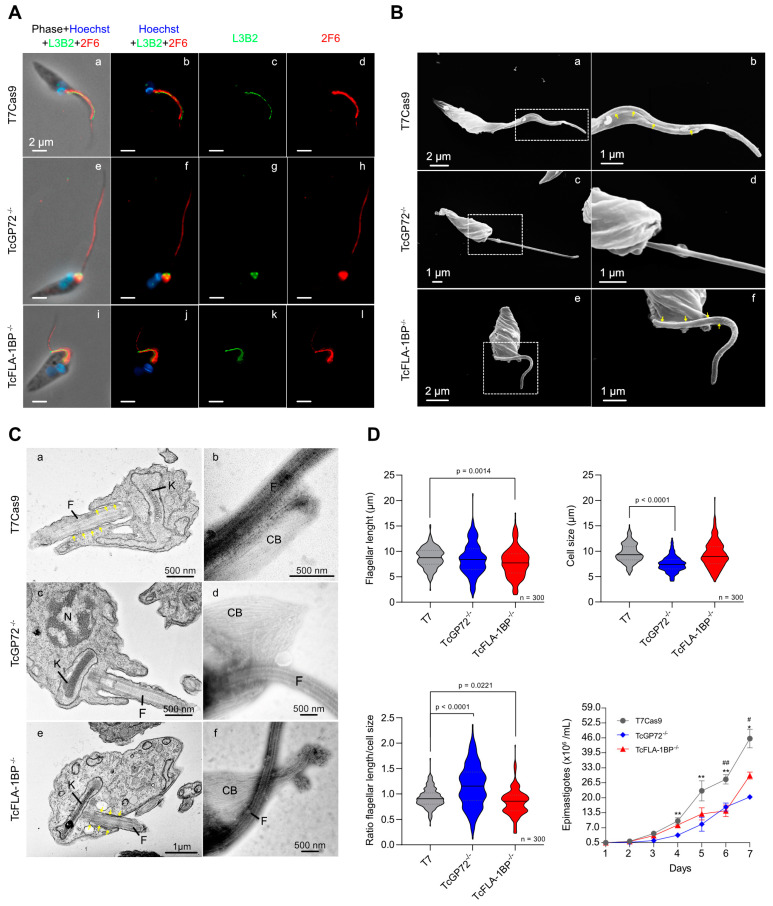
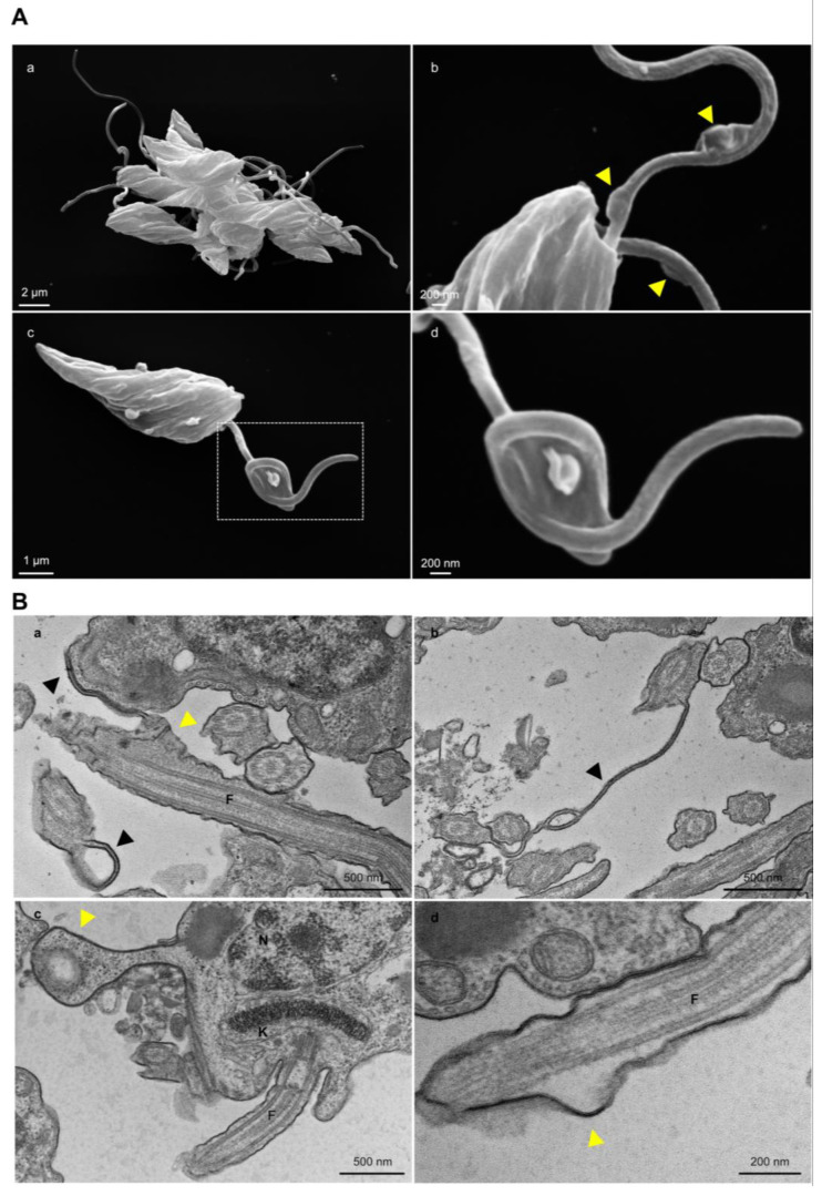
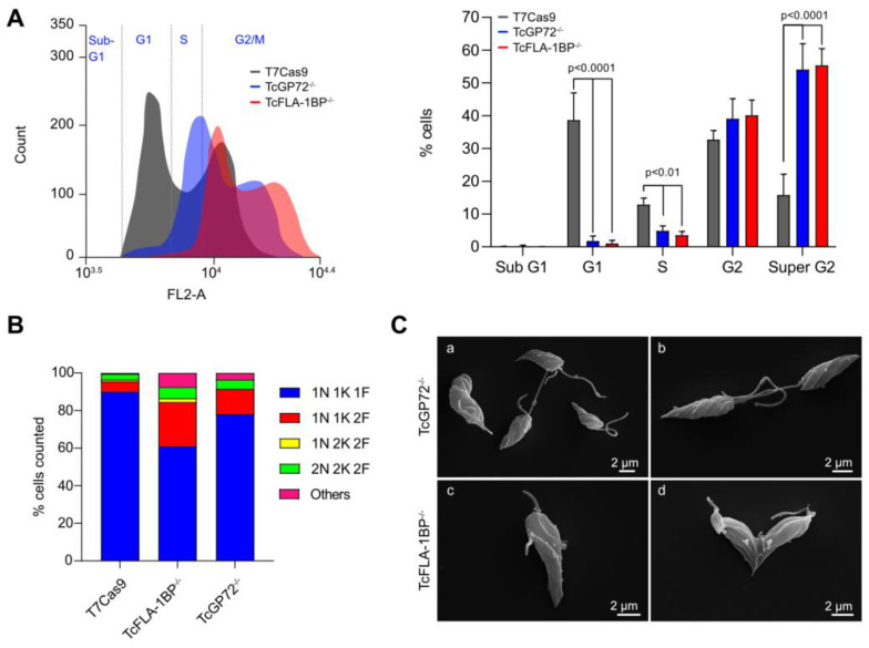
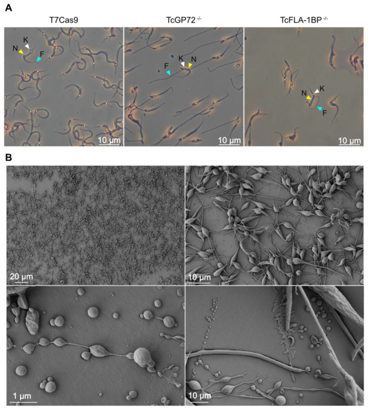
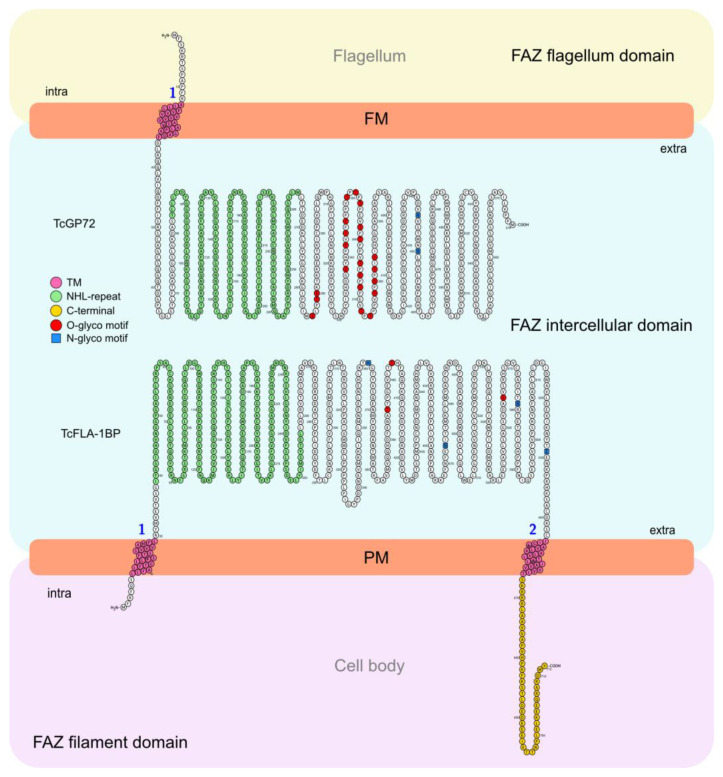
The highly adaptable parasite Trypanosoma cruzi undergoes complex developmental stages to exploit host organisms effectively. Each stage involves the expression of specific proteins and precise intracellular structural organization. These morphological changes depend on key structures that control intracellular components' growth and redistribution. In trypanosomatids, the flagellar attachment zone (FAZ) connects the flagellum to the cell body and plays a pivotal role in cell expansion and structural rearrangement. While FAZ proteins are well-studied in other trypanosomatids, there is limited knowledge about specific components, organization, and function in T. cruzi. This study employed the CRISPR/Cas9 system to label endogenous genes and conduct deletions to characterize FAZ-specific proteins during epimastigote cell division and metacyclogenesis. In T. cruzi, these proteins exhibited distinct organization compared to their counterparts in T. brucei. TcGP72 is anchored to the flagellar membrane, while TcFLA-1BP is anchored to the membrane lining the cell body. We identified unique features in the organization and function of the FAZ in T. cruzi compared to other trypanosomatids. Deleting these proteins had varying effects on intracellular structures, cytokinesis, and metacyclogenesis. This study reveals specific variations that directly impact the success of cell division and differentiation of this parasite.

Keywords: CRISPR/Cas9; flagellar attachment zone; morphological changes.


Conflict of interest statement

The authors declare no conflict of interest.

Figures

Figure 1
Figure 1
Bioinformatics analysis and domain organization of TcGP72 and TcFLA-1BP. (A) Phylogenetic tree from protein sequences of trypanosomatids. Amino acid sequences were aligned using Clustal Omega https://www.ebi.ac.uk/Tools/msa/clustalo/ (accessed on 22 April 2020, and the aligned sequences were used to generate a phylogenetic tree (SeaView 5.4). (B) Diagram of the conserved domains found in TcGP72 and TcFLA-1BP orthologous to Trypanosoma brucei. TcGP72 contains a transmembrane domain (TM) at the N-terminus and an NHL-repeat in the extracellular domain. TcFLA-1BP is structured with a transmembrane domain (TM) at the N-terminus, followed by NHL-repeat-containing extracellular domain, TM, and a 45-amino acid tail (C45) at the C-terminus. Numbers indicate the amino acid residues. The domains in the diagram were labeled based on the information deposited in the TritrypDB database. Domain organization is shown for TcGP72 and FLA-1BP fused to c-Myc and mNeonGreen tags at the C-terminus (3xMyc::mNG::3xMyc), named TcGP72::mNG and TcFLA-1BP::mNG. Tb: Trypanosoma brucei. Tc: Trypanosoma cruzi. Tev: Trypanosoma evansi. TvY486: Trypanosoma vivax. DQO04: Trypanosoma grayi. ADEAN: Angomonas deanei. CFAC1: Crithidia fasciculata. LbrM: Leishmania braziliensis. LAMA: Leishmania amazonensis. LINF: Leishmania infantum. LdCL: Leishmania donovani. LmjF: Leishmania major strain.
Figure 2
Figure 2
Analysis of the expression of fluorescent proteins. (A) Representative histograms showing the expression of fluorescent proteins in the mutant parasites. The peak with a black dashed line represents T7Cas9 lineage (control), and peaks with blue and red lines show TcGP72::mNG and TcFLA-1BP::mNG parasites, respectively. (B) Western blot analysis of fluorescent parasite (epimastigote) lysates expressing C-Myc and mNeonGreen fusion proteins (equivalent to 107 cells). Blots were probed with anti-Myc (AcMyc) antibodies and re-probed with anti-aldolase (AcAldo). kDa: molecular weight marker. The asterisk (*) shows the tagged protein bands. TcGP72::mNG: 98.2 kDa. TcFLA-1BP::mNG 116 kDa. TcAldolase: 29.3 kDa.
Figure 3
Figure 3
Localization of TcGP72::mNG and TcFLA-1BP::mNG proteins. (A) The fluorescent parasites generated with CRISPR/Cas9 were analyzed with fluorescence microscopy. Direct mNeonGreen fluorescence (green) reveals the cellular distribution of TcGP72::mNG (c,d) and TcFLA-1BP::mNG (e,f). TcGP72::mNG localized with distributed reticulated on the cell body and flagellum. In contrast, TcFLA-1BP::mNG was localized in just the FAZ. To confirm the FAZ domain localization of the TcFLA-1BP, we performed TcGP72 knockout in the TcFLA-1BP::mNG parasite (TcFLA-1BP::mNGΔGP72). TcFLA-1BP::mNGΔGP72 (g,h) parasites showed the detached flagellum and tagging exclusively on the side of the cell body along the FAZ. DAPI for DNA (blue). K: kinetoplast, N: nucleus. (B) Scanning electron microscopy shows TcGP72::mNG parasite (c,d) morphology with flagellum detachment in contrast to the T7Cas9 control parasite (a,b). The dotted region shows the magnified area, and the yellow arrows show the FAZ.
Figure 4
Figure 4
TcFLA-1BP targets the FAZ in amastigotes and trypomastigote (TCT) forms. (A) Fluorescence microscopy of TcFLA-1BP::mNG and T7Cas9 control in intracellular amastigotes (IAs) with FAZ labeled with anti-L3B2 (FAZ-1 T. brucei; red) and anti-mNG (mNeonGreen tag; green). (B) Localization in tissue culture derived TCTs. Nuclei and kinetoplasts were stained with Hoechst (blue). Scale bars: 2 and 5 µm.
Figure 5
Figure 5
Depletion of TcGP72 and TcFLA−1BP. Illustration of gene knockout strategy (not to scale). (A) Diagram showing the TcGP72 and TcFLA-1BP locus and PCR primers (arrows) used to confirm the presence of the TcGP72 and TcFLA-1BP coding sequence (blue box) or the correct integration of the drug-resistant genes (green box). An arrowhead marks Cas9 cleavage sites. Yellow and purple boxes represent the untranslated regions (UTRs) of endogenous genes and donor DNAs. (B,C) PCR products were visualized on agarose gel. T7Cas9, parental cell line; TcGP72−/−, double knockout parasite to TcGP72; TcGP72−/−, double knockout parasite to TcFLA-1BP. P1 (forward primer) anneals to the 5′ UTR of TcGP72 or TcFLA-1BP. P2 (reverse primer) anneals to the coding sequence of TcGP72 or TcFLA-1BP. P5 (reverse primer) anneals to the end of the resistance gene coding sequence (blasticidin or hygromicin).
Figure 6
Figure 6
TcFLA-1BP and TcGP72 knockout in T. cruzi causes partial or total flagellum detachment and impairs assembly of the flagellum, morphogenesis, organelle positioning, and cell growth. (A) Fluorescence microscopy of TcGP72::mNG and TcFLA-1BP::mNG with FAZ labeled with anti-L3B2 (FAZ-1 T. brucei; green) or anti-2F6 (PFR; red). Nuclei and kinetoplasts were stained with Hoechst (blue). Scale bars: 2 µm. T7Cas9 (ad). TcGP72−/− (eh). TcFLA-1BP−/− (il). (B) SEM showing the morphology of knockout parasites. TcGP72−/− (c,d) with total flagellum detachment and TcFLA-1BP−/− (e,f) partial detachment relative to the T7Cas9 control parasite (a,b). (C) TEM (a,c,e) and negative staining TEM (b,d,f) analysis. (a,c,e) Axial section of epimastigote forms showing the cell body (CB), kinetoplast (K), nuclei (N), flagellum (F), and FAZ (yellow arrows). (b,d,f) Negatively stained epimastigote forms showing the subpellicular microtubule of the cell body (CB) with a fully detached flagellum as in TcGP72−/− or a partially detached flagellum as in TcFLA-1BP−/−. (D) Violin plots show flagellum length, cell size, and the relation between flagellum length and cell size in the epimastigote forms. The mean values are indicated with a black line. Statistically significant differences are indicated with p-values. n = 300 cells. The line chart depicts the growth curves of the knockout parasites. Statistically significant differences are indicated with asterisks for TcGP72−/− and pound signs for TcFLA-1BP−/− in relation to the T7Cas9 control (* or # p < 0.05 and ** or ## p < 0.001).
Figure 7
Figure 7
TcGP72 knockout parasites display rosette formation, protrusions, and projection along the flagellum. (A) SEM of TcGP72−/− epimastigotes. (a) Rosette formation. (b) Flagellar membrane protrusions (yellow arrowhead) and (c,d) flagellar structures bizarrely filled with amorphous material. The dotted region (c) shows the magnified area (d). (B) TEM shows (a,b) the membrane projections (black arrowhead) and (a,c,d) protrusions (yellow arrowhead) of the flagellum and the cell body membrane in TcGP72 knockout parasites. F: Flagellum.
Figure 8
Figure 8
TcGP72 and TcFLA-1BP knockout in T. cruzi epimastigotes show cell cycle defects. Cell cycle comparative analysis with flow cytometry of the knockout parasites. (A) Representative histograms showing the DNA content of TcGP72−/−, TcFLA-1BP−/−, and the T7Cas9 control. The peak in gray represents the T7Cas9 parasites and peaks in red and blue represent depleted TcFLA-1BP and TcGP72, respectively. The bar graph represents the percentage of cells in each cell cycle phase in three independent experiments. Statistical analysis was performed with two-way ANOVA with Bonferroni correction for multiple testing. (B) Quantification of nuclei, the kinetoplast, and the flagellum in cells labeled for immunofluorescence microscopy with anti-2F6 (PFR) and Hoechst stain of knockout parasites (n = 300 cells). (C) SEM showing the abnormal morphology of knockout parasites. TcGP72−/− (a,b) and TcFLA-1BP−/− (c,d).
Figure 9
Figure 9
TcGP72 and TcFLA−1BP knockout T. cruzi show metacyclogenesis defects. (A) Metacyclic TCTs were purified on a DEAE cellulose column stained with Giemsa and observed under a microscope. The white arrowhead represents the kinetoplast, the yellow arrowhead represents the nucleus, and the blue arrowhead represents the flagellum. (B) Scanning microscopy images of TcGP72−/− parasites forming an adhesion network at the bottom of the culture flask during the metacyclogenesis assay.
Figure 10
Figure 10
A simplified view of the flagellum and cell body adhesion mediated by the interaction of TcGP72 and FLA-1BP in Trypanosoma cruzi. In T. cruzi, the flagellum membrane interacts with the cell body membrane, forming an adhesive network of proteins. This region is called the flagellar attachment zone (FAZ). The FAZ is divided into 3 main domains: (1) FAZ flagellar domain, (2) FAZ intercellular domain, and (3) FAZ filament domain. The components of the FAZ are distributed along the domains. Both TcGP72 and FLA1BP are essential for this adhesion at different intensities. In our model, TcGP72 is present on the flagellar membrane, and TcFLA-1BP is present on the cell membrane. TcGP72 is anchored to the flagellum by the intracellular domain at the N-terminus. TcFLA-1BP is anchored to the cell body by the two intracellular domains (N- and C-terminus). TcGP72 and TcFLA-1BP interact via the extracellular domains directed towards the intercellular domain of FAZ, thereby mediating flagellum/cell body adhesion. The numerals 1 and 2, highlighted in blue, denote the transmembrane regions. This interaction happens via the NHL-repeat domains present in the extracellular regions. The absence of TcGP72 and TcFLA-1BP causes partial or complete detachment of the flagellum. Both proteins have N- and O-glycosylation sites. Extra: extracellular. Intra: intracellular. FM: flagellum membrane. PM: plasma membrane.

Similar articles

Cited by

References

    1. Brener Z. Biology of Trypanosoma cruzi. Annu. Rev. Microbiol. 1973;27:347–382. doi: 10.1146/annurev.mi.27.100173.002023. - DOI - PubMed
    1. Burleigh B.A., Andrews N.W. The Mechanisms of Trypanosoma cruzi Invasion of Mammalian Cells. Annu. Rev. Microbiol. 1995;49:175–200. doi: 10.1146/annurev.mi.49.100195.001135. - DOI - PubMed
    1. De Souza W. International Review of Cytology. Volume 86. Academic Press; Cambridge, MA, USA: 1984. Cell Biology of Trypanosoma cruzi; pp. 197–283. - PubMed
    1. Fernandes M.C., Andrews N.W. Host Cell Invasion by Trypanosoma cruzi: A Unique Strategy That Promotes Persistence. FEMS Microbiol. Rev. 2012;36:734–747. doi: 10.1111/j.1574-6976.2012.00333.x. - DOI - PMC - PubMed
    1. Moretti N.S., Mortara R.A., Schenkman S. Trypanosoma cruzi. Trends Parasitol. 2020;36:404–405. doi: 10.1016/j.pt.2019.10.002. - DOI - PubMed

LinkOut - more resources