. 2023 Oct 30;13(11):1556.
doi: 10.3390/jpm13111556.

Immune Portrayal of a New Therapy Targeting Microbiota in an Animal Model of Psoriasis

Affiliations

Immune Portrayal of a New Therapy Targeting Microbiota in an Animal Model of Psoriasis

Mihaela Surcel et al. J Pers Med. .

Abstract

Background: Despite all the available treatments, psoriasis remains incurable; therefore, finding personalized therapies is a continuous challenge. Psoriasis is linked to a gut microbiota imbalance, highlighting the importance of the gut-skin axis and its inflammatory mediators. Restoring this imbalance can open new perspectives in psoriasis therapy. We investigated the effect of purified IgY raised against pathological human bacteria antibiotic-resistant in induced murine psoriatic dermatitis (PSO).

Methods: To evaluate the immune portrayal in an imiquimod experimental model, before and after IgY treatment, xMAP array and flow cytometry were used.

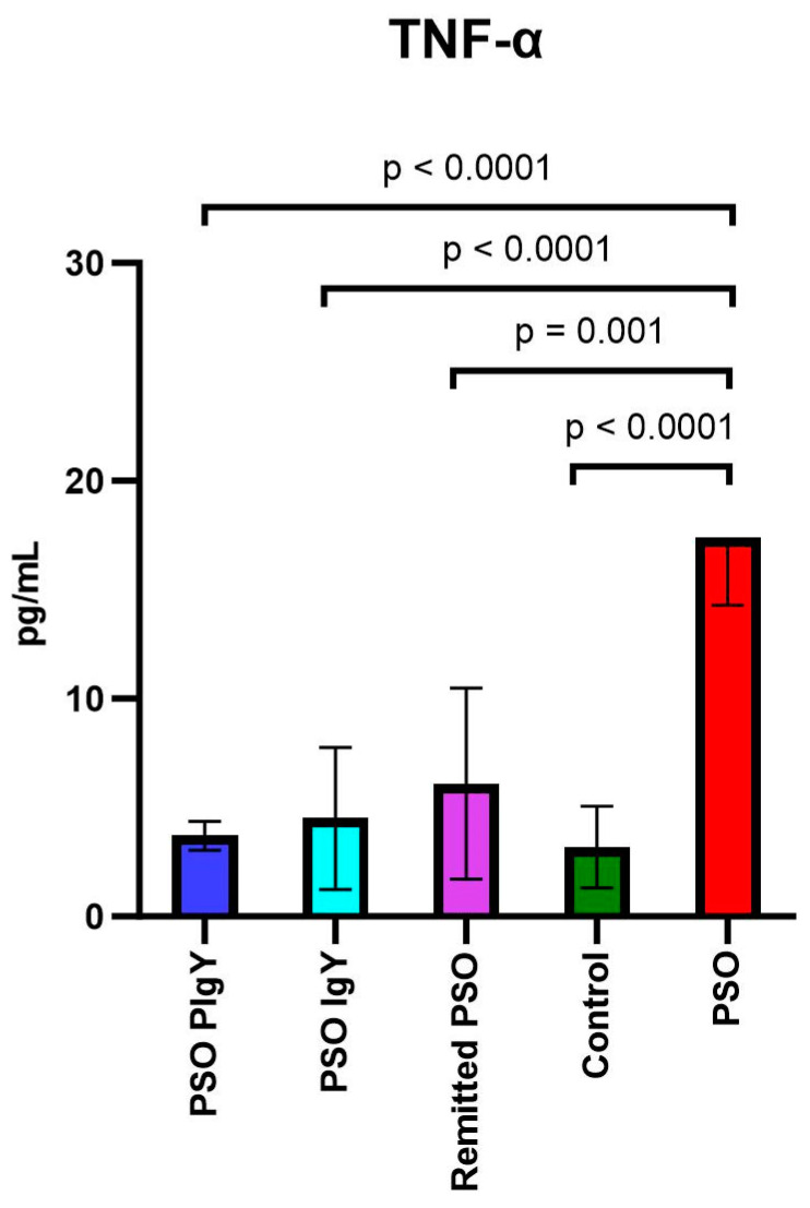
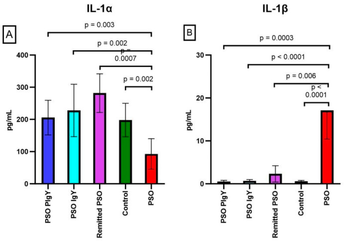
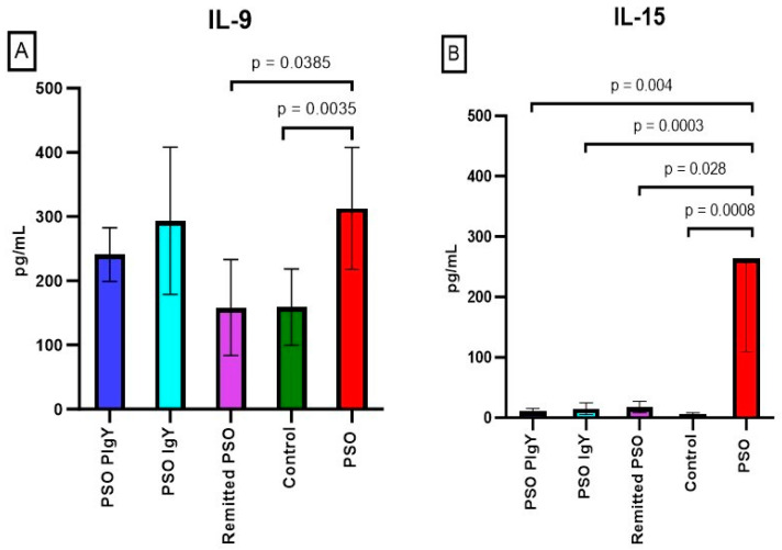
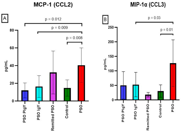
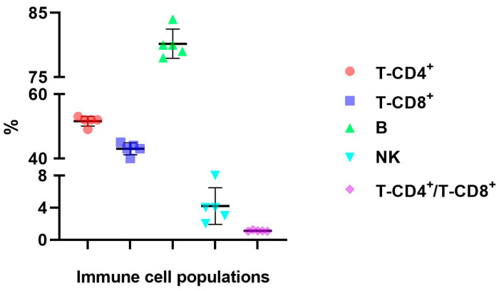
Results: There were significant changes in IL-1α,β, IL-5, IL-6, IL-9, IL-10, IL-12 (p70), IL-13, IL-15, IL-17a, IFN-γ, TNF-α, IP-10/CXCL10, MCP-1/CCL2, MIP-1α/CCL3, MIP-1β/CCL4, MIG/CXCL9, and KC/CXCL1 serum levels. T (CD3ε+), B (CD19+) and NK (NK1.1+) cells were also quantified. In our model, TNF-α, IL-6, and IL-1β cytokines and CXCL1 chemokine have extremely high circulatory levels in the PSO group. Upon experimental therapy, the cytokine serum values were not different between IgY-treated groups and spontaneously remitted PSO.

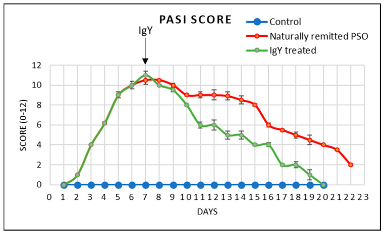
Conclusions: Using the murine model of psoriatic dermatitis, we show that the orally purified IgY treatment can lead to an improvement in skin lesion healing along with the normalization of cellular and humoral immune parameters.

Keywords: IgY; cytokines; gut–skin axis; inflammation; psoriatic dermatitis.


Conflict of interest statement

The authors declare no conflict of interest.

Figures

Figure 1
Figure 1
Parameters studied in the experimental murine model. IgY raised against pathological human bacteria resistant to antibiotics were orally administered in PSO-C57 BL/6 mice; cytokine/chemokine serum levels were followed in relation to the circulatory immune cell populations.
Figure 2
Figure 2
The study design over time. The graphic scheme of the study includes the main key points of the experiment: induction of murine PSO with IMQ (Day 0–Day 6), oral administration of IgY/PIgY treatment by gavage (Day 7–Day 11), healing period (up to Days 20/22), and harvesting of biological samples (Days 7, 20, and 22).
Figure 3
Figure 3
Gel filtration standard chromatogram for the optimization of sample preparation. Detection at 280 nm of thyroglobulin (Mr 670,000 Da), bovine γ-globulin (Mr 158,000 Da), chicken ovalbumin (Mr 44,000 Da), equine myoglobin (Mr 17,000 Da), and vitamin B12 (Mr 1350 Da).
Figure 4
Figure 4
IgY standard’s and IgY sample’s overlapped chromatograms. The IgY standard (yellow) and IgY sample chromatograms (blue) were obtained at a flow rate of 1 mL/min, 233 PSI, absorbance at 280 nm, and pH of 8.
Figure 5
Figure 5
PASI score in the control, in the naturally remitted PSO, and the IgY-treated groups (mean ± SD).
Figure 6
Figure 6
Serum levels of IL-12 (p70) and IL-6. (A). IL-12 (p70) in the PSO PIgY group (7.1 ± 3.6), PSO IgY group (8.8 ± 7.5), remitted PSO group (15.3 ± 1.4), control (6.3 ± 2.0), and untreated PSO group (50.3 ± 17.4); (B). IL-6 in the PSO PIgY group (4.2 ± 3.0), PSO IgY group (3.9 ± 5.5), remitted PSO group (10.8 ± 6.8), control (4.5 ± 3.3), and untreated PSO group (24.1 ± 6.2). The results are presented as the mean concentration values ± SD.
Figure 7
Figure 7
Serum levels of TNF-α. TNF-α in the PSO PIgY group (3.7 ± 0.7), PSO IgY group (4.5 ± 3.3), remitted PSO group (6.1 ± 4.4), control (3.2 ± 1.9), and untreated PSO group (17.4 ± 3.1). The results are presented as the mean concentration values ± SD.
Figure 8
Figure 8
Serum levels of IL-1α and IL-1β. (A) IL-1α in the PSO PIgY group (205.8 ± 53.9), PSO IgY group (227.8 ± 81.3), remitted PSO group (281.8 ± 59.9), control (198.1 ± 51.9), and PSO group (92.8 ± 47.5). (B) IL-1β in the PSO PIgY group (0.5 ± 0.3), PSO IgY group (0.7 ± 0.3), remitted PSO group (2.3 ± 1.9), control (0.6 ± 0.2), and PSO group (17.1 ± 6.7). The results are presented as the mean concentration values ± SD.
Figure 9
Figure 9
Serum levels of IL-9 and IL-15. (A) IL-9 in the PSO-PIgY group (241.0 ± 41.9), PSO IgY group (293.5 ± 114.5), remitted PSO group (158.3 ± 74.8), control (159.1 ± 59.3), and the untreated PSO group (312.5 ± 95.0). (B) IL-15 in the PSO PIgY group (10.5 ± 4.9), PSO IgY group (15.2 ± 9.7), remitted PSO group (17.9 ± 9.3), control (5.8 ± 2.5), and untreated PSO group (263.6 ± 154.4). The results are presented as the mean concentration values ± SD.
Figure 10
Figure 10
Serum levels of IL-5 and IL-10. (A) IL-5 in the PSO PIgY group (52.9 ± 6.0), PSO IgY group (56.5 ± 17.5), remitted PSO group (80.7 ± 28.6), control (33.7 ± 13.3), and untreated PSO group (180.3 ± 36.5). (B) IL-10 in the PSO PIgY group (1.7 ± 0.7), PSO IgY group (2.6 ± 2.1), remitted PSO group (4.1 ± 3.3), control (1.8 ± 1.3), and untreated PSO group (49.3 ± 18.7). The results are presented as the mean concentration values ± SD.
Figure 11
Figure 11
Serum levels of MCP-1/CCL2 and MIP-1α/CCL3. (A) MCP-1/CCL2 in the PSO PIgY group (12.0 ± 8.4), PSO IgY group (16.3 ± 12.4), remitted PSO group (32.2 ± 24.3), control (14.7 ± 9.2), and untreated PSO group (40.5 ± 19.6). (B) MIP-1α/CCL3 in the PSO PIgY group (50.2 ± 47.3), PSO IgY group (52.7 ± 42.2), remitted PSO group (18.1 ± 7.9), control (30.6 ± 21.4), and untreated PSO group (126.5 ± 80.4). The results are presented as the mean concentration values ± SD.
Figure 12
Figure 12
Serum levels of KC/CXCL1, IP-10/CXCL10, and MIG/CXCL9. (A) KC/CXCL1 in the PSO PIgY group (153.7 ± 41.0), PSO IgY group (161.4 ± 32.1), remitted PSO group (223.2 ± 57.8), control (161.0 ± 76.5), and untreated PSO group (232.0 ± 22.6). (B) IP-10/CXCL10 in the PSO PIgY group (297.1 ± 83.7), PSO IgY group (283.1 ± 66.3), remitted PSO group (316.1 ± 71.5), control (278.4 ± 47.6), and untreated PSO group (346.7 ± 52.6). (C) Expression of MIG/CXCL9 in the PSO PIgY group (252.2 ± 59.9), PSO IgY group (373.8 ± 92.9), remitted PSO group (396.3 ± 107.2), control (282.3 ± 85.5), and untreated PSO group (474.9 ± 61.9). The results are presented as the mean concentration values ± SD.
Figure 13
Figure 13
Distribution of the immune cell populations in the peripheral blood harvested from purified IgY-treated mice. Distribution of T-CD4+ (52 ± 1.4) and T-CD8+ (43 ± 1.9) lymphocyte subsets, B cells (80 ± 3.2), NK cells (4 ± 2.1), and T-CD4+/T-CD8+ ratio (1.20 ± 0.1) in the PSO PIgY group. The results are presented as mean values ± SD.

Similar articles

Cited by

References

    1. Tashiro T., Sawada Y. Psoriasis and Systemic Inflammatory Disorders. Int. J. Mol. Sci. 2022;23:4457. doi: 10.3390/ijms23084457. - DOI - PMC - PubMed
    1. Jansen J.E., Aschenbrenner D., Uhlig H.H., Coles M.C., Gaffney E.A. A method for the inference of cytokine interaction networks. PLoS Comput. Biol. 2022;18:e1010112. doi: 10.1371/journal.pcbi.1010112. - DOI - PMC - PubMed
    1. Lee H.R., Sung J.H. Multiorgan-on-a-chip for realization of gut-skin axis. Biotechnol. Bioeng. 2022;119:2590–2601. doi: 10.1002/bit.28164. - DOI - PubMed
    1. Ion A., Dorobanțu A.M., Popa L.G., Mihai M.M., Orzan O.A. Risks of Biologic Therapy and the Importance of Multidisciplinary Approach for an Accurate Management of Patients with Moderate-Severe Psoriasis and Concomitant Diseases. Biology. 2022;11:808. doi: 10.3390/biology11060808. - DOI - PMC - PubMed
    1. Moludi J., Fathollahi P., Khedmatgozar H., Pourteymour Fard Tabrizi F., Ghareaghaj Zare A., Razmi H. Probiotics Supplementation Improves Quality of Life, Clinical Symptoms, and Inflammatory Status in Patients with Psoriasis. J. Drugs Dermatol. 2022;21:637–644. doi: 10.36849/JDD.6237. - DOI - PubMed