

Retinal Findings and Cardiovascular Risk: Prognostic Conditions, Novel Biomarkers, and Emerging Image Analysis Techniques
- PMID: 38003879
- PMCID: PMC10672409
- DOI: 10.3390/jpm13111564
Retinal Findings and Cardiovascular Risk: Prognostic Conditions, Novel Biomarkers, and Emerging Image Analysis Techniques
Abstract
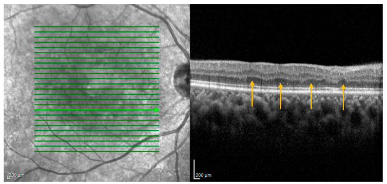
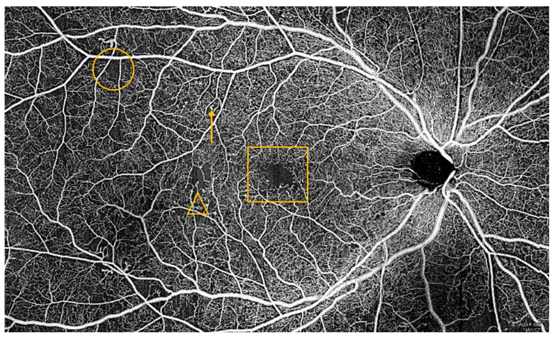
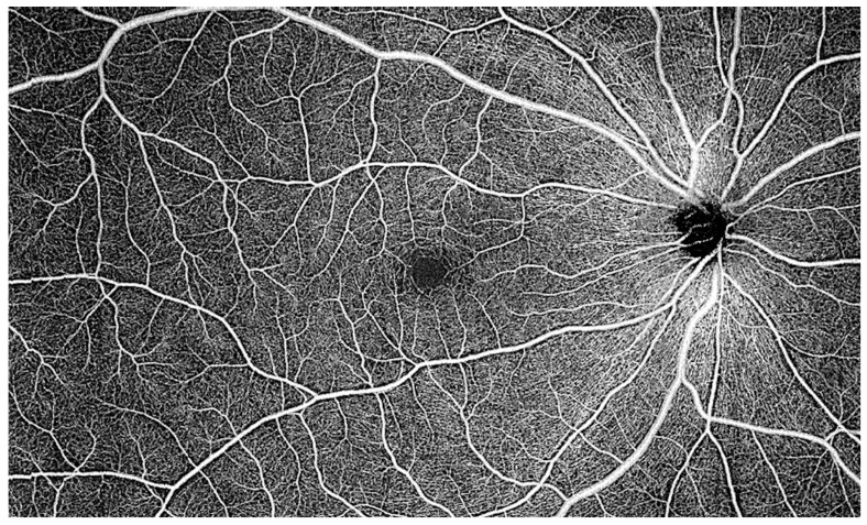
Many retinal diseases and imaging findings have pathophysiologic underpinnings in the function of the cardiovascular system. Myriad retinal conditions, new imaging biomarkers, and novel image analysis techniques have been investigated for their association with future cardiovascular risk or utility in cardiovascular risk prognostication. An intensive literature search was performed to identify relevant articles indexed in PubMed, Scopus, and Google Scholar for a targeted narrative review. This review investigates the literature on specific retinal disease states, such as retinal arterial and venous occlusions and cotton wool spots, that portend significantly increased risk of future cardiovascular events, such as stroke or myocardial infarction, and the implications for personalized patient counseling. Furthermore, conditions diagnosed primarily through retinal bioimaging, such as paracentral acute middle maculopathy and the newly discovered entity known as a retinal ischemic perivascular lesion, may be associated with future incident cardiovascular morbidity and are also discussed. As ever-more-sophisticated imaging biomarkers and analysis techniques are developed, the review concludes with a focused analysis of optical coherence tomography and optical coherence tomography angiography biomarkers under investigation for potential value in prognostication and personalized therapy in cardiovascular disease.
Keywords: cardiovascular risk; myocardial infarction; optical coherence tomography; optical coherence tomography angiography; retinal ischemic perivascular lesions; retinal vascular occlusions; stroke; subretinal drusenoid deposits.
Conflict of interest statement
The authors declare no conflict of interests.
Figures
Similar articles
-
Retinal Ischemic Perivascular Lesions (RIPLs) as Potential Biomarkers for Systemic Vascular Diseases: A Narrative Review of the Literature.Ophthalmol Ther. 2025 Jun;14(6):1183-1197. doi: 10.1007/s40123-025-01148-5. Epub 2025 Apr 28. Ophthalmol Ther. 2025. PMID: 40293678 Free PMC article. Review.
-
Retinal vascular occlusion in pregnancy: three case reports and a review of the literature.J Med Case Rep. 2022 Apr 21;16(1):167. doi: 10.1186/s13256-022-03369-9. J Med Case Rep. 2022. PMID: 35449024 Free PMC article. Review.
-
Differences in the multicolour imaging features between the superficial and deep vascular occlusions.Int Ophthalmol. 2020 Dec;40(12):3431-3439. doi: 10.1007/s10792-020-01529-8. Epub 2020 Jul 31. Int Ophthalmol. 2020. PMID: 32737726
-
SUPERFICIAL AND DEEP CAPILLARY ISCHEMIA AS A PRESENTING SIGN OF RETINAL VASCULOPATHY WITH CEREBRAL LEUKOENCEPHALOPATHY AND SYSTEMIC MANIFESTATIONS.Retin Cases Brief Rep. 2018 Fall;12 Suppl 1:S87-S91. doi: 10.1097/ICB.0000000000000641. Retin Cases Brief Rep. 2018. PMID: 29028736
-
Paracentral acute middle maculopathy spectral-domain optical coherence tomography feature of deep capillary ischemia.Curr Opin Ophthalmol. 2014 May;25(3):207-12. doi: 10.1097/ICU.0000000000000045. Curr Opin Ophthalmol. 2014. PMID: 24614148 Review.
Cited by
-
Retinal Imaging as a Window into Cardiovascular Health: Towards Harnessing Retinal Analytics for Precision Cardiovascular Medicine.J Cardiovasc Dev Dis. 2025 Jun 17;12(6):230. doi: 10.3390/jcdd12060230. J Cardiovasc Dev Dis. 2025. PMID: 40558665 Free PMC article. Review.
-
Exploring Endothelial Cell Dysfunction's Impact on the Brain-Retina Microenvironment Connection: Molecular Mechanisms and Implications.Mol Neurobiol. 2025 Jun;62(6):7484-7505. doi: 10.1007/s12035-025-04714-x. Epub 2025 Feb 4. Mol Neurobiol. 2025. PMID: 39904964 Review.
-
Optical coherence tomography angiography in cardiovascular disease.Prog Cardiovasc Dis. 2024 Nov-Dec;87:60-72. doi: 10.1016/j.pcad.2024.10.011. Epub 2024 Oct 21. Prog Cardiovasc Dis. 2024. PMID: 39442597 Review.
-
Comparison of Vascular Density Changes After Cataract Surgery in Diabetic Patients with and Without Pseudoexfoliation Syndrome Using Optical Coherence Tomography Angiography.Biomedicines. 2025 Apr 8;13(4):908. doi: 10.3390/biomedicines13040908. Biomedicines. 2025. PMID: 40299487 Free PMC article.
References
-
- Mac Grory B., Schrag M., Biousse V., Furie K.L., Gerhard-Herman M., Lavin P.J., Sobrin L., Tjoumakaris S.I., Weyand C.M., Yaghi S. Management of Central Retinal Artery Occlusion: A Scientific Statement from the American Heart Association. Stroke. 2021;52:e282–e294. doi: 10.1161/STR.0000000000000366. - DOI - PubMed
Publication types
LinkOut - more resources
Full Text Sources
Research Materials
