Adipose-Derived Mesenchymal Stem Cell (MSC) Immortalization by Modulation of hTERT and TP53 Expression Levels
- PMID: 38003936
- PMCID: PMC10672200
- DOI: 10.3390/jpm13111621
Adipose-Derived Mesenchymal Stem Cell (MSC) Immortalization by Modulation of hTERT and TP53 Expression Levels
Abstract
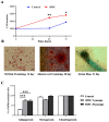
Mesenchymal stem cells (MSCs) are pivotal players in tissue repair and hold great promise as cell therapeutic agents for regenerative medicine. Additionally, they play a significant role in the development of various human diseases. Studies on MSC biology have encountered a limiting property of these cells, which includes a low number of passages and a decrease in differentiation potential during in vitro culture. Although common methods of immortalization through gene manipulations of cells are well established, the resulting MSCs vary in differentiation potential compared to primary cells and eventually undergo senescence. This study aimed to immortalize primary adipose-derived MSCs by overexpressing human telomerase reverse transcriptase (hTERT) gene combined with a knockdown of TP53. The research demonstrated that immortalized MSCs maintained a stable level of differentiation into osteogenic and chondrogenic lineages during 30 passages, while also exhibiting an increase in cell proliferation rate and differentiation potential towards the adipogenic lineage. Long-term culture of immortalized cells did not alter cell morphology and self-renewal potential. Consequently, a genetically stable line of immortalized adipose-derived MSCs (iMSCs) was established.
Keywords: MSC; MSC differentiation; immortalization; mesenchymal stem cell; tumor stroma.
Conflict of interest statement
The authors declare no conflict of interest.
Figures


Similar articles
-
Generation of Mesenchymal Cell Lines Derived from Aged Donors.Int J Mol Sci. 2021 Oct 1;22(19):10667. doi: 10.3390/ijms221910667. Int J Mol Sci. 2021. PMID: 34639008 Free PMC article.
-
Molecular basis of immortalization of human mesenchymal stem cells by combination of p53 knockdown and human telomerase reverse transcriptase overexpression.Stem Cells Dev. 2013 Jan 15;22(2):268-78. doi: 10.1089/scd.2012.0222. Epub 2012 Aug 21. Stem Cells Dev. 2013. PMID: 22765508 Free PMC article.
-
Deer antler stem cells immortalization by modulation of hTERT and the small extracellular vesicles characters.Front Vet Sci. 2024 Oct 4;11:1440855. doi: 10.3389/fvets.2024.1440855. eCollection 2024. Front Vet Sci. 2024. PMID: 39430380 Free PMC article.
-
Meta-analysis of the Mesenchymal Stem Cells Immortalization Protocols: A Guideline for Regenerative Medicine.Curr Stem Cell Res Ther. 2024;19(7):1009-1020. doi: 10.2174/011574888X268464231016070900. Curr Stem Cell Res Ther. 2024. PMID: 38221663
-
Immortalized cell lines derived from dental/odontogenic tissue.Cell Tissue Res. 2023 Jul;393(1):1-15. doi: 10.1007/s00441-023-03767-5. Epub 2023 Apr 11. Cell Tissue Res. 2023. PMID: 37039940 Review.
Cited by
-
Advances in the Toxicity Assessment of Silver Nanoparticles Derived from a Sphagnum fallax Extract for Monolayers and Spheroids.Biomolecules. 2024 May 22;14(6):611. doi: 10.3390/biom14060611. Biomolecules. 2024. PMID: 38927015 Free PMC article.
-
In Vitro Analysis of PMEPA1 Upregulation in Mesenchymal Stem Cells Induced by Prostate Cancer Cells.Int J Mol Sci. 2025 Jun 27;26(13):6223. doi: 10.3390/ijms26136223. Int J Mol Sci. 2025. PMID: 40649997 Free PMC article.
-
Optimization of 3D Extrusion-Printed Particle-Containing Hydrogels for Osteogenic Differentiation.ACS Omega. 2025 Apr 10;10(15):15036-15051. doi: 10.1021/acsomega.4c10515. eCollection 2025 Apr 22. ACS Omega. 2025. PMID: 40290951 Free PMC article.
-
Multicellular Cancer-Stroma Spheres (CSS) for In Vitro Assessment of CAR-T Cell-Associated Toxicity.Cells. 2024 Nov 16;13(22):1892. doi: 10.3390/cells13221892. Cells. 2024. PMID: 39594640 Free PMC article.
References
-
- Witwer K.W., Van Balkom B.W.M., Bruno S., Choo A., Dominici M., Gimona M., Hill A.F., De Kleijn D., Koh M., Lai R.C., et al. Defining mesenchymal stromal cell (MSC)-derived small extracellular vesicles for therapeutic applications. J. Extracell. Vesicles. 2019;8:1609206. doi: 10.1080/20013078.2019.1609206. - DOI - PMC - PubMed
-
- Dominici M., Le Blanc K., Mueller I., Slaper-Cortenbach I., Marini F., Krause D., Deans R., Keating A., Prockop D., Horwitz E. Minimal criteria for defining multipotent mesenchymal stromal cells. The International Society for Cellular Therapy position statement. Cytotherapy. 2006;8:315–317. doi: 10.1080/14653240600855905. - DOI - PubMed
Grants and funding
LinkOut - more resources
Full Text Sources
Research Materials
Miscellaneous

