

Introducing an Innovative Approach for Managing Proximal Non-Cavitated Carious Lesions in Juvenile Permanent Dentition: Combining Orthodontic Separators and Silver Fluoride Application
- PMID: 38003942
- PMCID: PMC10672833
- DOI: 10.3390/medicina59111892
Introducing an Innovative Approach for Managing Proximal Non-Cavitated Carious Lesions in Juvenile Permanent Dentition: Combining Orthodontic Separators and Silver Fluoride Application
Abstract
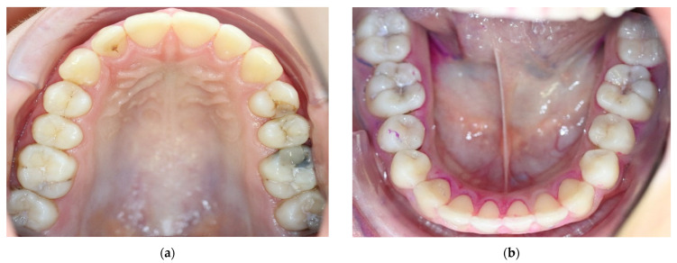
Background and objectives: The aim was to introduce an innovative, easy and cheap clinical approach for the control of multiple proximal non-cavitated lesions via the application of 38% silver fluoride after placement of orthodontic separators in the permanent dentition in high-caries-risk children.
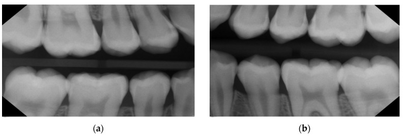
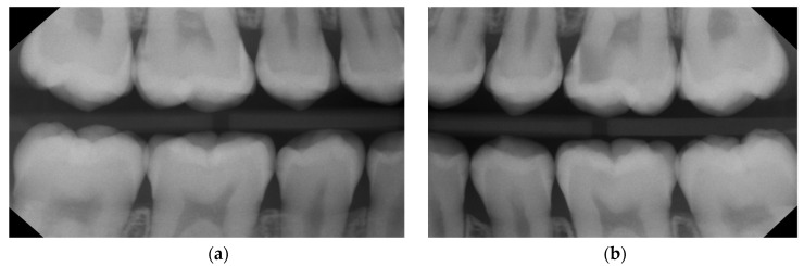
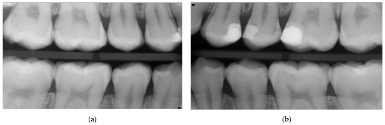
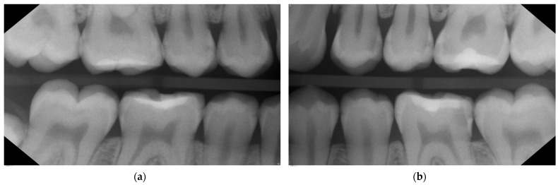
Materials and methods: The case series describes the management of initial proximal carious lesions using silver fluoride (SF) products in the permanent dentition of two adolescent patients with prior proximal caries progression. Both presented with multiple asymptomatic carious lesions that were identified through the use of bitewing radiographs and classified according to the ADA proximal caries classification system. Using orthodontic separators prior to the planned application of SF, most of the surfaces could then be quickly directly examined to check for surface integrity. Follow-up examinations were conducted clinically and radiographically for at least one year to monitor lesion progression.
Results: None of the 25 enamel lesions (E1/E2) exhibited signs of progression after single SF application, while 2 out of 11 dentine lesions (D1) showed progression and required restorative intervention. The progressed lesions potentially had non-cleansable micro-cavitations that were either clinically undetected or not fully reached with the micro-brush in SF application. Thus, this should have been repeated or combined with SF application via soaked superfloss to potentially achieve better results.
Conclusions: Single application of 38% silver fluoride directly onto active enamel lesions in juvenile permanent teeth with the prior use of orthodontic separators combined with a caries-risk-specific prevention program appears to be highly effective and should be considered as a viable minimally invasive option for patients and clinicians due to its cost-effectiveness and time efficiency.
Keywords: caries diagnosis; initial caries; minimal invasive; silver fluoride.
Conflict of interest statement
The authors declare no personal conflict of interest and no external funding for this project. It is worthwhile mentioning that the department of preventive and pediatric dentistry at the university medicine Greifswald has received partial funding from the company SDI for conducting other scientific projects.
Figures


Similar articles
-
When to intervene in the caries process? An expert Delphi consensus statement.Clin Oral Investig. 2019 Oct;23(10):3691-3703. doi: 10.1007/s00784-019-03058-w. Epub 2019 Aug 23. Clin Oral Investig. 2019. PMID: 31444695
-
Silver Diamine Fluoride and Progression of Incipient Approximal Caries in Permanent Teeth: A Retrospective Study.Pediatr Dent. 2021 Nov 15;43(6):475-480. Pediatr Dent. 2021. PMID: 34937619
-
Silver diamine fluoride remineralized artificial incipient caries in permanent teeth after bacterial pH-cycling in-vitro.J Dent. 2018 Feb;69:55-59. doi: 10.1016/j.jdent.2017.09.005. Epub 2017 Sep 14. J Dent. 2018. PMID: 28918101
-
Prevention of Dental Caries by Silver Diamine Fluoride.Compend Contin Educ Dent. 2019 Mar;40(3):158-163; quiz 164. Compend Contin Educ Dent. 2019. PMID: 30829497 Review.
-
How to intervene in the caries process: proximal caries in adolescents and adults-a systematic review and meta-analysis.Clin Oral Investig. 2020 May;24(5):1623-1636. doi: 10.1007/s00784-020-03201-y. Epub 2020 Apr 18. Clin Oral Investig. 2020. PMID: 32306093
Cited by
-
Updates on Caries Management in the Primary and Permanent Dentition.Medicina (Kaunas). 2025 Feb 11;61(2):316. doi: 10.3390/medicina61020316. Medicina (Kaunas). 2025. PMID: 40005433 Free PMC article.
-
Caries Lesion Assessment Using 3D Virtual Models by Examiners with Different Degrees of Clinical Experience.Medicina (Kaunas). 2023 Dec 13;59(12):2157. doi: 10.3390/medicina59122157. Medicina (Kaunas). 2023. PMID: 38138260 Free PMC article.
References
-
- Rechmann P., Kinsel R., Featherstone J.D.B. Integrating Caries Management by Risk Assessment (CAMBRA) and Prevention Strategies Into the Contemporary Dental Practice. Compend. Contin. Educ. Dent. 2018;39:226. - PubMed
-
- Agarwal D., Machale P.S., Hegde-Shetiya S. The Incipient Caries. J. Contemp. Dent. 2013;3:20–24. doi: 10.5005/jp-journals-10031-1029. - DOI
Publication types
MeSH terms
Substances
LinkOut - more resources
Full Text Sources
Medical
Research Materials
