

The Impact of the Addition of Vitamins on a Silicone Lining Material to the Oral Mucosa Tissue-Evaluation of the Biocompatibility, Hydrolytic Stability and Histopathological Effect
- PMID: 38003985
- PMCID: PMC10673301
- DOI: 10.3390/medicina59111936
The Impact of the Addition of Vitamins on a Silicone Lining Material to the Oral Mucosa Tissue-Evaluation of the Biocompatibility, Hydrolytic Stability and Histopathological Effect
Abstract
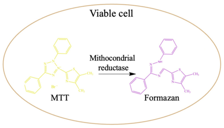
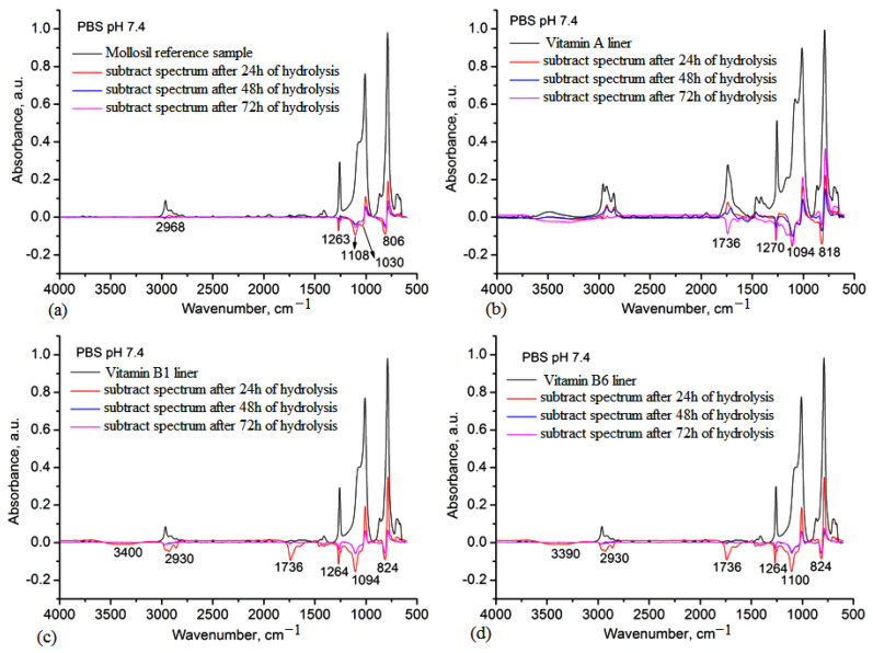
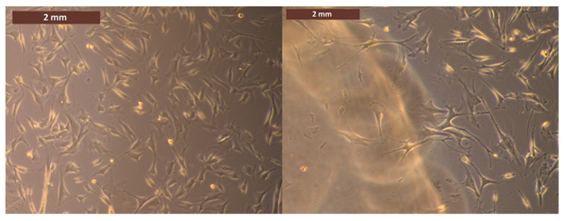
Background and Objectives: One's quality of life depends on overall health, and in particular, oral health, which has been and continues to become a public health issue through frequent manifestations in various forms, from simple oral stomatitis (inflammations of the oral cavity) to the complicated oral health pathologies requiring medical interventions and treatments (caries, pulp necrosis and periodontitis). The aim of this study focused on the preparation and evaluation of vitamins (vitamin A, B1 and B6) incorporated into several silicone-based lining materials as a new alternative to therapeutically loaded materials designed as oral cavity lining materials in prosthodontics. Materials and Methods: Silicone-based liners containing vitamins were prepared by mixing them in solution and becoming crosslinked, and then they were characterized using Fourier-transform infrared (FT-IR) spectroscopy to confirm the incorporation of the vitamins into the silicone network; scanning electron microscopy (SEM) to evidence the morphology of the liner materials; dynamic vapor sorption (DVS) to evaluate their internal hydrophobicity, swelling in environments similar to biological fluids and mechanical test to demonstrate tensile strength; MTT to confirm their biocompatibility on normal cell cultures (fibroblast) and mucoadhesivity; and histopathological tests on porcine oral mucosa to highlight their potential utility as soft lining materials with improved efficiency. Results: FT-IR analysis confirmed the structural peculiarities of the prepared lining materials and the successful incorporation of vitamins into the silicone matrix. The surface roughness of the materials was lower than 0.2 μm, while in cross-section, the lining materials showed a compact morphology. It was found that the presence of vitamins induced a decrease in the main mechanical parameters (strength and elongation at break, Young's modulus) and hydrophobicity, which varied from one vitamin to another. A swelling degree higher than 8% was found in PBS 6.8 (artificial saliva) and water. Hydrolytic stability studies in an artificial saliva medium showed the release of low concentrations of silicone and vitamin fragments in the first 24 h, which increased the swelling behavior of the materials, diffusion and solubility of the vitamins. The microscopic images of fibroblast cells incubated with vitamin liners revealed very good biocompatibility. Also, the silicone liners incorporating the vitamins showed good mucoadhesive properties. The appearance of some pathological disorders with autolysis processes was more pronounced in the case of vitamin A liners. Conclusions: The addition of the vitamins was shown to have a beneficial effect that was mainly manifested as increased biocompatibility, hydrolytic stability and mucoadhesiveness with the mucosa of the oral cavity and less of an effect on the mechanical strength. The obtained lining materials showed good resistance in simulated biological media but caused a pronounced autolysis phenomenon, as revealed by histopathological examination, showing that these materials may have broad implications in the treatment of oral diseases.
Keywords: biocompatibity; histopathology; lining materials; oral health; stability in biological media; vitamins.
Conflict of interest statement
The authors declare no conflict of interest.
Figures


Similar articles
-
The effect of surface roughness of silicone-based resilient liner materials on the adherence of Candida albicans and inhibition of Candida albicans with different disinfectants.Oral Health Prev Dent. 2009;7(4):347-53. Oral Health Prev Dent. 2009. PMID: 20011752
-
Effect of thermocycling on tensile bond strength of six silicone-based, resilient denture liners.J Prosthet Dent. 2003 Mar;89(3):303-10. doi: 10.1067/mpr.2003.41. J Prosthet Dent. 2003. PMID: 12644808
-
Effect of storage duration on tensile bond strength of acrylic or silicone-based soft denture liners to a processed denture base polymer.Acta Odontol Scand. 2005 Feb;63(1):31-5. doi: 10.1080/00016350510019667. Acta Odontol Scand. 2005. PMID: 16095060
-
Advances in Soft Denture Liners: An Update.J Contemp Dent Pract. 2015 Apr 1;16(4):314-8. doi: 10.5005/jp-journals-10024-1682. J Contemp Dent Pract. 2015. PMID: 26067736 Review.
-
Soft lining materials--a review.Eur J Prosthodont Restor Dent. 1995 Jun;3(4):163-74. Eur J Prosthodont Restor Dent. 1995. PMID: 8601159 Review.
References
MeSH terms
Substances
LinkOut - more resources
Full Text Sources
