

Giant Mandibular Ameloblastoma with Rare Hypercalcemia: A Case Report and Literature Review
- PMID: 38004005
- PMCID: PMC10673442
- DOI: 10.3390/medicina59111956
Giant Mandibular Ameloblastoma with Rare Hypercalcemia: A Case Report and Literature Review
Abstract
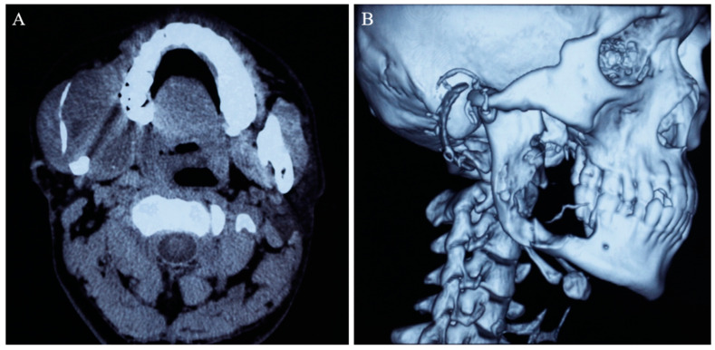
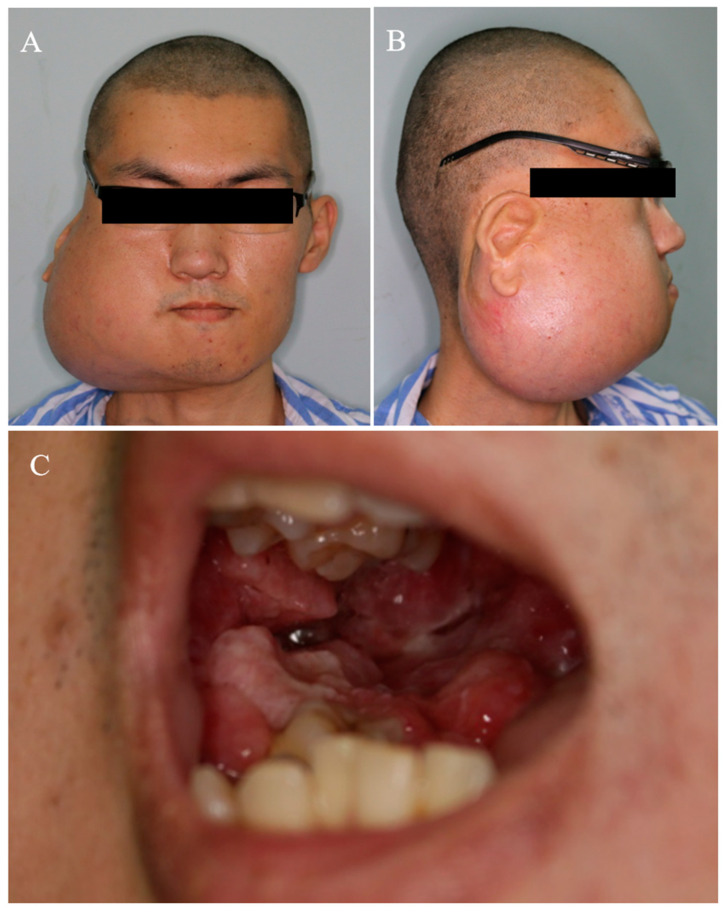
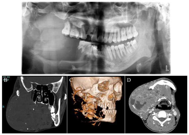
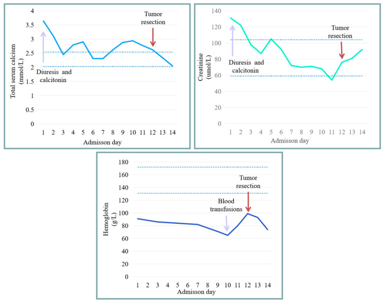
Ameloblastoma is the most common benign odontogenic tumor with local invasion and high recurrence, which generally occurs in the jaw bones. Hypercalcemia is a common paraneoplastic syndrome that is commonly observed in patients with malignancies but rarely encountered in patients with benign tumors. Thus far, not many cases of ameloblastoma with hypercalcemia have been reported, and the pathogenic mechanism has not been studied in depth. This paper presents a case report of a 26-year-old male diagnosed with giant ameloblastoma of the mandible, accompanied by rare hypercalcemia. Additionally, a review of the relevant literature is conducted. This patient initially underwent marsupialization, yet this treatment was not effective, which indicated that the selection of the appropriate operation is of prime importance for improving the prognosis of patients with ameloblastoma. The tumor not only failed to shrink but gradually increased in size, accompanied by multiple complications including hypercalcemia, renal dysfunction, anemia, and cachexia. Due to the contradiction between the necessity of tumor resection and the patient's poor systemic condition, we implemented a multi-disciplinary team (MDT) meeting to better evaluate this patient's condition and design an individualized treatment strategy. The patient subsequently received a variety of interventions to improve the general conditions until he could tolerate surgery, and finally underwent the successful resection of giant ameloblastoma and reconstruction with vascularized fibular flap. No tumor recurrence or distance metastasis was observed during 5 years of follow-up. Additionally, the absence of hypercalcemia recurrence was also noted.
Keywords: ameloblastoma; hypercalcemia; marsupialization; multi-disciplinary teams; renal impairment; surgery.
Conflict of interest statement
The authors declare no conflict of interest.
Figures
Similar articles
-
Overwhelming hypercalcaemia in mandibular ameloblastoma.BMJ Case Rep. 2014 Oct 17;2014:bcr2014205491. doi: 10.1136/bcr-2014-205491. BMJ Case Rep. 2014. PMID: 25326561 Free PMC article.
-
Ameloblastic carcinoma ex ameloblastoma of the mandible with malignancy-associated hypercalcemia.Oral Surg Oral Med Oral Pathol Oral Radiol Endod. 2000 Dec;90(6):716-22. doi: 10.1067/moe.2000.109076. Oral Surg Oral Med Oral Pathol Oral Radiol Endod. 2000. PMID: 11113817
-
Recurrence of ameloblastoma in bone grafts of a fibula free flap: A case report and literature review.J Stomatol Oral Maxillofac Surg. 2022 Nov;123(6):663-665. doi: 10.1016/j.jormas.2022.06.007. Epub 2022 Jun 10. J Stomatol Oral Maxillofac Surg. 2022. PMID: 35697254 Review.
-
Giant ameloblastoma: report of an extreme case and a description of its treatment.Ear Nose Throat J. 1999 Aug;78(8):568, 570-2, 574. Ear Nose Throat J. 1999. PMID: 10485149
-
Pulmonary metastasectomy in the treatment of recurrent ameloblastoma of the maxilla and mandible: a case report.Eur Arch Otorhinolaryngol. 2001 Jan;258(1):25-7. doi: 10.1007/s004050000293. Eur Arch Otorhinolaryngol. 2001. PMID: 11271430 Review.
References
Publication types
MeSH terms
Grants and funding
LinkOut - more resources
Full Text Sources
