

A Rare Case of Isolated Hepatocellular Carcinoma Metastasis in Left Mandibular Region in a Patient with Hepatitis C Virus Liver Cirrhosis Diagnosed after the Onset of COVID-19 Infection
- PMID: 38004041
- PMCID: PMC10673151
- DOI: 10.3390/medicina59111992
A Rare Case of Isolated Hepatocellular Carcinoma Metastasis in Left Mandibular Region in a Patient with Hepatitis C Virus Liver Cirrhosis Diagnosed after the Onset of COVID-19 Infection
Abstract
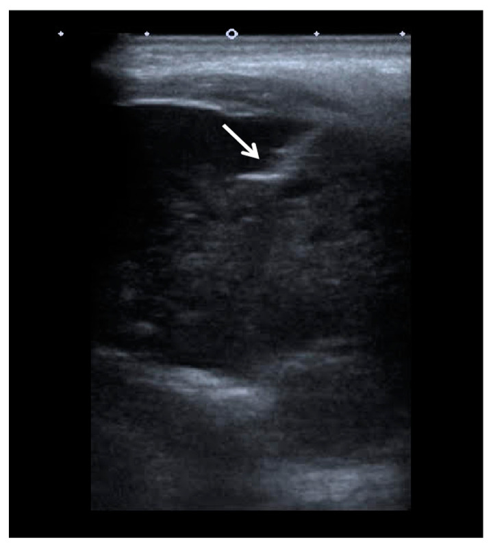
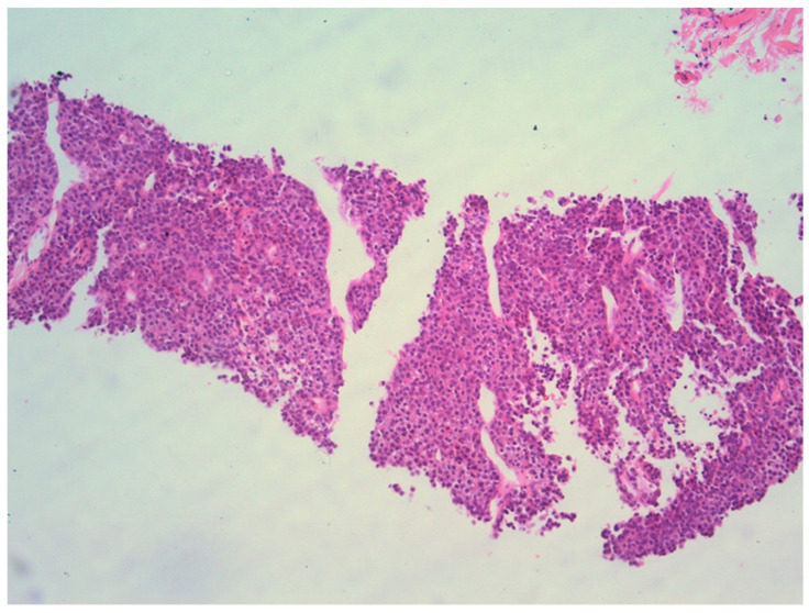
Background and Objectives: Hepatocellular carcinoma (HCC) most frequently metastasizes in the lungs, abdominal lymph nodes and adrenal glands. Metastatic spread to the head and neck area is extremely rare. In the presented case, an uncommon site of solitary metastatic spread of HCC to the mandible confirmed after the core biopsy of the lesion is reported. There have been only about 80 cases of mandibular HCC metastases described in the literature to date. We contribute our experience to the pool of data. Case presentation: A 65-year-old female with HCV-related liver cirrhosis was diagnosed with an HCC that was successfully treated with liver resection. Subsequently, the patient had developed COVID-19 disease, which was associated with a painless swelling in the left jaw. A neck MDCT scan demonstrated an osteolytic soft-tissue mass in the left mandible, with the characteristics consistent for the metastasis of HCC. In order to confirm the diagnosis, a core biopsy of the mandibular mass was performed. The pathohistological evaluation confirmed the presence of a metastatic HCC in the mandible. No other sites of disease dissemination were identified in extensive MDCT scans. Despite considering various treatments, including symptomatic and palliative, the patient's overall prognosis remained poor. Conclusions: Isolated metastases of HCC to the orofacial region are extremely rare; however, it should be considered in patients with known risk factors for HCC development. Early diagnosis is critical, and clinicians should consider this possibility of HCC spread when assessing patients with orofacial swelling, among those patients with risk factors for HCC. The overall prognosis for such patients remains poor, emphasizing the challenges in managing these cases.
Keywords: biopsy; diagnosis; hepatocellular carcinoma; mandible; metastasis.
Conflict of interest statement
The authors declare no conflict of interest.
Figures


Similar articles
-
Unresectable hepatocellular carcinoma with a solitary metastasis to the mandible.Am Surg. 2008 Apr;74(4):346-9. Am Surg. 2008. PMID: 18453303
-
Intractable bleeding from solitary mandibular metastasis of hepatocellular carcinoma.World J Gastroenterol. 2007 Sep 7;13(33):4526-8. doi: 10.3748/wjg.v13.i33.4526. World J Gastroenterol. 2007. PMID: 17724815 Free PMC article.
-
Solitary mandibular metastasis as an initial manifestation of hepatocellular carcinoma.Acta Med Okayama. 2006 Aug;60(4):243-7. doi: 10.18926/AMO/30713. Acta Med Okayama. 2006. PMID: 16943863
-
Metastasis to the jaws as a first manifestation of hepatocellular carcinoma: report of a case and analysis of 41 cases.J Craniomaxillofac Surg. 2014 Dec;42(8):1997-2001. doi: 10.1016/j.jcms.2014.09.005. Epub 2014 Sep 19. J Craniomaxillofac Surg. 2014. PMID: 25441863 Review.
-
Gingival metastasis from primary hepatocellular carcinoma: a case report and literature review of 30 cases.BMC Cancer. 2019 Sep 14;19(1):925. doi: 10.1186/s12885-019-6020-7. BMC Cancer. 2019. PMID: 31521125 Free PMC article. Review.
Cited by
-
Unusual extrahepatic metastatic site of hepatocellular carcinoma with post-therapy disseminating metastases presenting as a primary soft tissue sarcoma: case report.Ann Med Surg (Lond). 2024 Jun 25;86(9):5501-5508. doi: 10.1097/MS9.0000000000002307. eCollection 2024 Sep. Ann Med Surg (Lond). 2024. PMID: 39239049 Free PMC article.
References
Publication types
MeSH terms
LinkOut - more resources
Full Text Sources
Medical
