

Regeneration of Intrabony Defects Using a Novel Magnesium Membrane
- PMID: 38004067
- PMCID: PMC10672749
- DOI: 10.3390/medicina59112018
Regeneration of Intrabony Defects Using a Novel Magnesium Membrane
Abstract
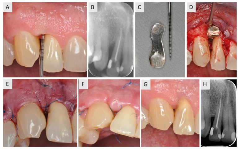
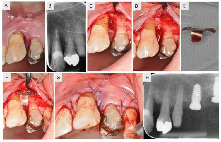
Background and Objectives: Due to their specific morphology, the regeneration of intrabony defects (IBDs) represents one of the greatest challenges for clinicians. Based on the specific properties of a magnesium membrane, a new approach for the surgical treatment of IBD was developed. The surgical procedure was described using a series of three cases. Materials and Methods: The patients were healthy individuals suffering from a severe form of periodontitis associated with IBD. Based on radiographic examination, the patients had interproximal bone loss of at least 4 mm. Due to its good mechanical properties, it was easy to cut and shape the magnesium membrane into three different shapes to treat the specific morphology of each IBD. In accordance with the principles of guided bone regeneration, a bovine xenograft was used to fill the IBD in all cases. Results: After a healing period of 4 to 6 months, successful bone regeneration was confirmed using radiological analysis. The periodontal probing depth (PPD) after healing showed a reduction of 1.66 ± 0.29 mm. Conclusions: Overall, the use of the different shapes of the magnesium membrane in the treatment of IBD resulted in a satisfactory functional and esthetic outcome.
Keywords: NOVAMag membrane; intrabony defects; magnesium; resorbable metal.
Conflict of interest statement
P.R., S.R. and Ž.P.K. are employees of botiss biomaterials GmbH.
Figures

Similar articles
-
Five-year results of guided tissue regeneration in combination with deproteinized bovine bone (Bio-Oss) in the treatment of intrabony periodontal defects: a case series report.Clin Oral Investig. 2005 Dec;9(4):271-7. doi: 10.1007/s00784-005-0002-7. Epub 2005 Jul 12. Clin Oral Investig. 2005. PMID: 16010581
-
Use of bovine-derived anorganic bone associated with guided tissue regeneration in intrabony defects. Six-month evaluation at re-entry.J Periodontol. 1999 Sep;70(9):1000-7. doi: 10.1902/jop.1999.70.9.1000. J Periodontol. 1999. PMID: 10505802 Clinical Trial.
-
Platelet-rich plasma and bovine porous bone mineral combined with guided tissue regeneration in the treatment of intrabony defects in humans.J Periodontal Res. 2002 Aug;37(4):300-6. doi: 10.1034/j.1600-0765.2002.01001.x. J Periodontal Res. 2002. PMID: 12200975 Clinical Trial.
-
Periodontal regeneration - intrabony defects: a consensus report from the AAP Regeneration Workshop.J Periodontol. 2015 Feb;86(2 Suppl):S105-7. doi: 10.1902/jop.2015.140378. Epub 2014 Oct 15. J Periodontol. 2015. PMID: 25315019
-
Osteogenic Potential of Magnesium Oxide Nanoparticles in Bone Regeneration: A Systematic Review.Cureus. 2024 Mar 4;16(3):e55502. doi: 10.7759/cureus.55502. eCollection 2024 Mar. Cureus. 2024. PMID: 38571856 Free PMC article. Review.
Cited by
-
Guided Bone Regeneration in the Posterior Mandible Using a Resorbable Metal Magnesium Membrane and Fixation Screws: A Case Report.Case Rep Dent. 2024 Dec 13;2024:2659893. doi: 10.1155/crid/2659893. eCollection 2024. Case Rep Dent. 2024. PMID: 39713245 Free PMC article.
-
Biomaterial Scaffolds for Periodontal Tissue Engineering.J Funct Biomater. 2024 Aug 20;15(8):233. doi: 10.3390/jfb15080233. J Funct Biomater. 2024. PMID: 39194671 Free PMC article. Review.
-
Soft Tissue Graft Placement Using a Porcine Acellular Dermal Matrix (PADM) and Resorbable Magnesium Fixation Screws: A Case Series.Medicina (Kaunas). 2025 Jun 25;61(7):1144. doi: 10.3390/medicina61071144. Medicina (Kaunas). 2025. PMID: 40731774 Free PMC article.
-
Implantable Dental Barrier Membranes as Regenerative Medicine in Dentistry: A Comprehensive Review.Tissue Eng Regen Med. 2025 Jun;22(4):527-549. doi: 10.1007/s13770-025-00704-1. Epub 2025 Feb 24. Tissue Eng Regen Med. 2025. PMID: 39992621 Free PMC article. Review.
References
-
- Azzi R., Takei H., Etienne D., Carranza F.A. Root coverage and papilla reconstruction using autogenous osseous and connective tissue grafts. Int. J. Periodontics Restor. Dent. 2001;21:141–147. - PubMed
MeSH terms
Substances
LinkOut - more resources
Full Text Sources
