. 2023 Nov 8;15(22):4722.
doi: 10.3390/nu15224722.

Effect of Extracelluar Vesicles Derived from Akkermansia muciniphila on Intestinal Barrier in Colitis Mice

Affiliations

Effect of Extracelluar Vesicles Derived from Akkermansia muciniphila on Intestinal Barrier in Colitis Mice

Ting Zheng et al. Nutrients. .

Abstract

Inflammatory bowel disease (IBD) is a chronic and recurrent disease. It has been observed that the incidence and prevalence of IBD are increasing, which consequently raises the risk of developing colon cancer. Recently, the regulation of the intestinal barrier by probiotics has become an effective treatment for colitis. Akkermansia muciniphila-derived extracellular vesicles (Akk EVs) are nano-vesicles that contain multiple bioactive macromolecules with the potential to modulate the intestinal barrier. In this study, we used ultrafiltration in conjunction with high-speed centrifugation to extract Akk EVs. A lipopolysaccharide (LPS)-induced RAW264.7 cell model was established to assess the anti-inflammatory effects of Akk EVs. It was found that Akk EVs were able to be absorbed by RAW264.7 cells and significantly reduce the expression of nitric oxide (NO), TNF-α, and IL-1β (p < 0.05). We explored the preventative effects on colitis and the regulating effects on the intestinal barrier using a mouse colitis model caused by dextran sulfate sodium (DSS). The findings demonstrated that Akk EVs effectively prevented colitis symptoms and reduced colonic tissue injury. Additionally, Akk EVs significantly enhanced the effectiveness of the intestinal barrier by elevating the expression of MUC2 (0.53 ± 0.07), improving mucus integrity, and reducing intestinal permeability (p < 0.05). Moreover, Akk EVs increased the proportion of the beneficial bacteria Firmicutes (33.01 ± 0.09%) and downregulated the proportion of the harmful bacteria Proteobacteria (0.32 ± 0.27%). These findings suggest that Akk EVs possess the ability to regulate immune responses, protect intestinal barriers, and modulate the gut microbiota. The research presents a potential intervention approach for Akk EVs to prevent colitis.

Keywords: Akkermansia muciniphila; colitis; extracellular vesicles; gut microbiota; intestinal barrier; prevention.


Conflict of interest statement

The authors have no competing interests to declare. All authors give their approval for the publication of this manuscript.

Figures

Figure 1
Figure 1
Characterization of Akk EVs. (a) TEM images of Akk EVs. (b) Size distribution of Akk EVs.
Figure 2
Figure 2
Akk EVs suppress LPS-induced inflammatory responses in RAW264.7 cells. (a) Fluorescence images of cell internalization profiles of Dil-loaded EVs in RAW264.7 cells. Scale bar represents 20 μm. Effects of Akk EVs on the production of NO (b), IL-1β (c), and TNF-α (d) in the presence of 100 ng/mL LPS in RAW264.7 cells. All data were presented as mean ± SD (n = 5). * p < 0.05, compared with the LPS group; # p < 0.05, compared with the control group.
Figure 3
Figure 3
Experimental schedule and basic indicators. (a) Animal treatment schedule. (b) Bodyweight change in each group. (c) DAI scores in each group. (d) Representative images of the colon length. (e) Colon length. All data were presented as mean ± SD (n = 6). Control group (Con), DSS group (DSS), Akk group (Akk), and Akk EVs group (EVs). * p < 0.05, compared with the DSS group; # p < 0.05, compared with the control group.
Figure 4
Figure 4
Histopathological Characteristics of colitis mice. (a) Typical histological sections stained with HE. (b) The scoring of colon histological injury. Scale bar represents 100 μm. Red arrows indicated colon lesions. Control group (Con), DSS group (DSS), Akk group (Akk), and Akk EVs group (EVs). * p < 0.05, compared with the DSS group; # p < 0.05, compared with the control group.
Figure 5
Figure 5
AB-PAS staining. (a) Typical histological sections stained with AB-PAS. (b) Mucin area (%). Scale bar represents 100 μm. Red arrows indicated acidic mucin and green arrows indicated mixed mucin. Control group (Con), DSS group (DSS), Akk group (Akk), and Akk EVs group (EVs). # p < 0.05, compared with the control group.
Figure 6
Figure 6
Effects of Akk EVs on immunoglobulins and inflammatory cytokine. (a) IgA. (b) sIgA. (c) MPO. (d) TNF-α. (e) IL-1β. (f) IL-6. All data were presented as mean ± SD (n = 6). Control group (Con), DSS group (DSS), Akk group (Akk), and Akk EVs group (EVs). * p < 0.05, compared with the DSS group; # p < 0.05, compared with the control group.
Figure 7
Figure 7
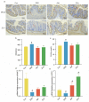
Effects of Akk EVs on intestinal permeability and barrier function. (a) Images of immunohistochemistry of MUC2 and ZO-1. Scale bar represents 100 μm. (b) Concentration of DAO. (c) Concentration of D-LA. (d) mRNA expression of MUC2. (e) mRNA expression of ZO-1. All data were presented as mean ± SD (n = 5). Red arrows indicated protein positive expression. Control group (Con), DSS group (DSS), Akk group (Akk), and Akk EVs group (EVs). * p < 0.05, compared with the DSS group; # p < 0.05, compared with the control group.
Figure 8
Figure 8
Effects of Akk EVs supplements on the gut microbiome structure. (a) Venn diagrams of OTUs. (b) Rarefaction curves. (c) Chao1 index. (d) PCoA analysis. (e) phylum level. (f) genus level. All data were presented as mean ± SD (n = 5). Control group (Con), DSS group (DSS), Akk group (Akk), and Akk EVs group (EVs). # p < 0.05, compared with the control group.
Figure 9
Figure 9
The effect of Akk EVs on gut dominant microorganisms. (a) The taxonomic tree of differentially abundant taxa based on genus was depicted by the cladogram. (b) Histogram of the distribution of dominant gut microorganisms at the genus level. Control group (Con), DSS group (DSS), Akk group (Akk), and Akk EVs group (EVs).
Figure 10
Figure 10
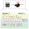
Schematic illustration of Akk EVs regulating intestinal barrier.

References

    1. Franzosa E.A., Sirota-Madi A., Avila-Pacheco J., Fornelos N., Haiser H.J., Reinker S., Vatanen T., Hall A.B., Mallick H., McIver L.J., et al. Gut Microbiome Structure and Metabolic Activity in Inflammatory Bowel Disease. Nat. Microbiol. 2018;4:293–305. doi: 10.1038/s41564-018-0306-4. - DOI - PMC - PubMed
    1. Kaplan G.G., Windsor J.W. The Four Epidemiological Stages in the Global Evolution of Inflammatory Bowel Disease. Nat. Rev. Gastroenterol. Hepatol. 2021;18:56–66. doi: 10.1038/s41575-020-00360-x. - DOI - PMC - PubMed
    1. Dong L., Xie J., Wang Y., Jiang H., Chen K., Li D., Wang J., Liu Y., He J., Zhou J., et al. Mannose Ameliorates Experimental Colitis by Protecting Intestinal Barrier Integrity. Nat. Commun. 2022;13:4804. doi: 10.1038/s41467-022-32505-8. - DOI - PMC - PubMed
    1. Halfvarson J., Brislawn C.J., Lamendella R., Vázquez-Baeza Y., Walters W.A., Bramer L.M., D’Amato M., Bonfiglio F., McDonald D., Gonzalez A., et al. Dynamics of the Human Gut Microbiome in Inflammatory Bowel Disease. Nat. Microbiol. 2017;2:17004. doi: 10.1038/nmicrobiol.2017.4. - DOI - PMC - PubMed
    1. Lee S.H. Intestinal Permeability Regulation by Tight Junction: Implication on Inflammatory Bowel Diseases. Intest. Res. 2015;13:11. doi: 10.5217/ir.2015.13.1.11. - DOI - PMC - PubMed

Substances

Supplementary concepts

LinkOut - more resources