Analysis of Kinematic and Muscular Fatigue in Long-Distance Swimmers
- PMID: 38004269
- PMCID: PMC10671841
- DOI: 10.3390/life13112129
Analysis of Kinematic and Muscular Fatigue in Long-Distance Swimmers
Abstract
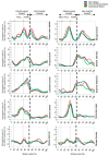
Muscle fatigue is a complex phenomenon that is influenced by the type of activity performed and often manifests as a decline in motor performance (mechanical failure). The purpose of our study was to investigate the compensatory strategies used to mitigate mechanical failure. A cohort of 21 swimmers underwent a front-crawl swimming task, which required the consistent maintenance of a constant speed for the maximum duration. The evaluation included three phases: non-fatigue, pre-mechanical failure, and mechanical failure. We quantified key kinematic metrics, including velocity, distance travelled, stroke frequency, stroke length, and stroke index. In addition, electromyographic (EMG) metrics, including the Root-Mean-Square amplitude and Mean Frequency of the EMG power spectrum, were obtained for 12 muscles to examine the electrical manifestations of muscle fatigue. Between the first and second phases, the athletes covered a distance of 919.38 ± 147.29 m at an average speed of 1.57 ± 0.08 m/s with an average muscle fatigue level of 12%. Almost all evaluated muscles showed a significant increase (p < 0.001) in their EMG activity, except for the latissimus dorsi, which showed a 17% reduction (ES 0.906, p < 0.001) during the push phase of the stroke cycle. Kinematic parameters showed a 6% decrease in stroke length (ES 0.948, p < 0.001), which was counteracted by a 7% increase in stroke frequency (ES -0.931, p < 0.001). Notably, the stroke index also decreased by 6% (ES 0.965, p < 0.001). In the third phase, characterised by the loss of the ability to maintain the predetermined rhythm, both EMG and kinematic parameters showed reductions compared to the previous two phases. Swimmers employed common compensatory strategies for coping with fatigue; however, the ability to maintain a predetermined motor output proved to be limited at certain levels of fatigue and loss of swimming efficiency (Protocol ID: NCT06069440).
Keywords: electromyography; muscle coordination; swimming; task failure.
Conflict of interest statement
The authors declare no conflict of interest.
Figures


Similar articles
-
Kinematic and electromyographic changes during 200 m front crawl at race pace.Int J Sports Med. 2013 Jan;34(1):49-55. doi: 10.1055/s-0032-1321889. Epub 2012 Aug 17. Int J Sports Med. 2013. PMID: 22903317
-
Kinematic and electromyography characteristics of performing butterfly stroke with different swimming speeds in flow environment.Heliyon. 2023 Sep 13;9(9):e20122. doi: 10.1016/j.heliyon.2023.e20122. eCollection 2023 Sep. Heliyon. 2023. PMID: 37809614 Free PMC article. Review.
-
Evaluation of muscle fatigue during 100-m front crawl.Eur J Appl Physiol. 2011 Jan;111(1):101-13. doi: 10.1007/s00421-010-1624-2. Epub 2010 Sep 8. Eur J Appl Physiol. 2011. PMID: 20824283
-
Surface Electromyography Spectral Parameters for the Study of Muscle Fatigue in Swimming.Front Sports Act Living. 2021 Feb 19;3:644765. doi: 10.3389/fspor.2021.644765. eCollection 2021. Front Sports Act Living. 2021. PMID: 33681763 Free PMC article.
-
How do swimmers control their front crawl swimming velocity? Current knowledge and gaps from hydrodynamic perspectives.Sports Biomech. 2023 Dec;22(12):1552-1571. doi: 10.1080/14763141.2021.1959946. Epub 2021 Aug 23. Sports Biomech. 2023. PMID: 34423742 Review.
Cited by
-
Biomechanical coordination and variability alters following repetitive movement fatigue in overhead athletes with painful shoulder.Sci Rep. 2025 Jan 3;15(1):718. doi: 10.1038/s41598-025-85226-5. Sci Rep. 2025. PMID: 39753727 Free PMC article.
References
Associated data
LinkOut - more resources
Full Text Sources
Medical

