

Improving Chest Monitoring through Magnetic Resonance Angiogram Image Contrast Enhancement
- PMID: 38004300
- PMCID: PMC10672579
- DOI: 10.3390/life13112160
Improving Chest Monitoring through Magnetic Resonance Angiogram Image Contrast Enhancement
Abstract
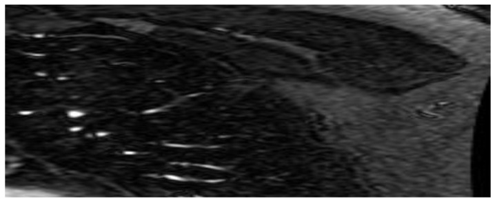
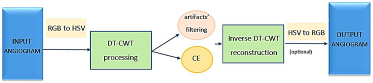
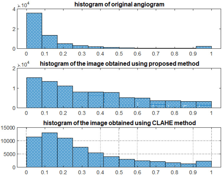
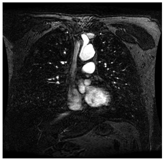
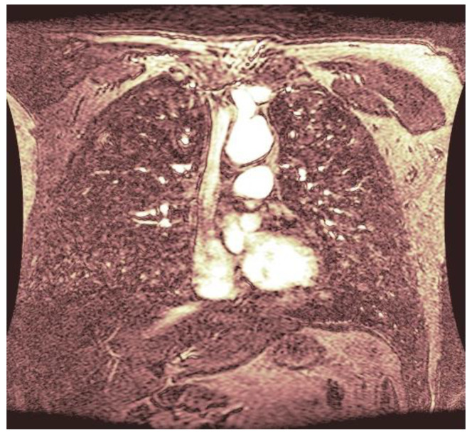
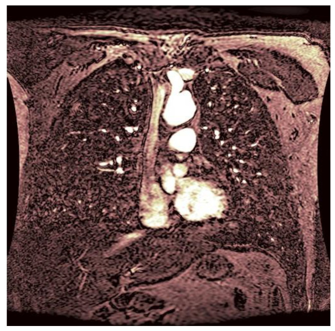
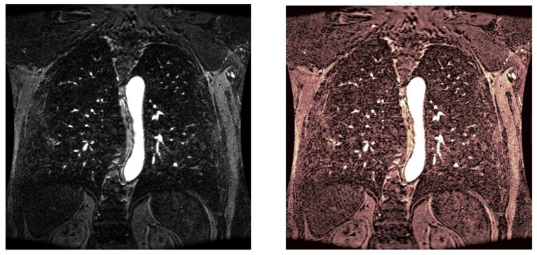
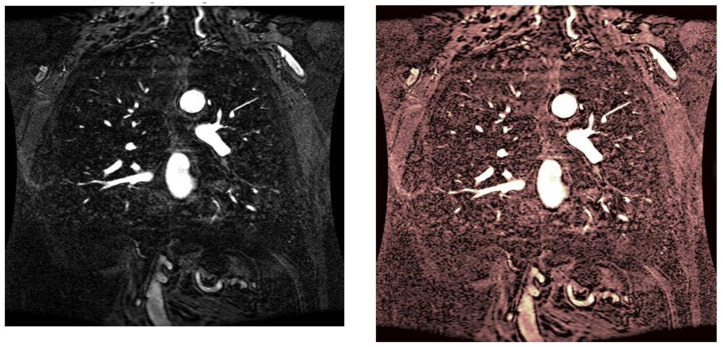
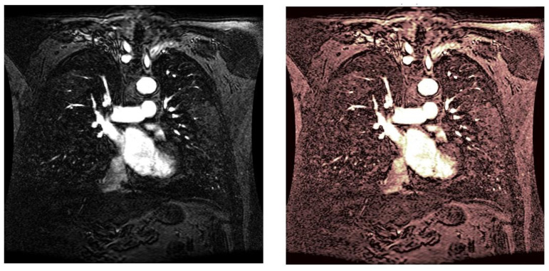
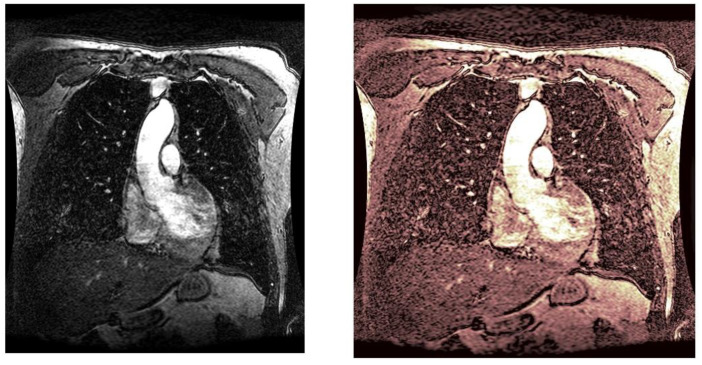
Magnetic resonance angiography is a medical procedure used to offer an image of the blood vessels and organs of the body. Given the worldwide spread of cardiovascular diseases, more and more resources are invested in treating them. One of the most modern treatments involves the acquisition of images of the heart. Sometimes the contrast of these images is not satisfactory. Injecting invasive enhancement substances to obtain a better view of the cardiac route is not advisable. However, software algorithms can solve the problem. This study proposes and tests a local adaptive contrast-adjustment algorithm using the dual-tree complex wavelet transform. The method has been tested with medical data from a public database to allow comparisons to other methods. The selected algorithm further improved the contrast of images. The performances are given for evaluation, both visually (to help doctors make accurate diagnoses) and in parametric form (to show engineers which parts of the algorithm might need improvement). Compared to other contrast enhancement methods, the proposed wavelet algorithm shows good results and greater stability. Thus, we aim to avoid future pointless complications due to unnecessary contrast substances.
Keywords: angiography; dual-tree discrete wavelet transform; filters.
Conflict of interest statement
The authors declare no conflict of interest.
Figures
Similar articles
-
Dual tree complex wavelet transform based denoising of optical microscopy images.Biomed Opt Express. 2012 Dec 1;3(12):3231-9. doi: 10.1364/BOE.3.003231. Epub 2012 Nov 13. Biomed Opt Express. 2012. PMID: 23243573 Free PMC article.
-
A wavelet-based method for improving signal-to-noise ratio and contrast in MR images.Magn Reson Imaging. 2000 Feb;18(2):169-80. doi: 10.1016/s0730-725x(99)00128-9. Magn Reson Imaging. 2000. PMID: 10722977
-
Non-linear direct multi-scale image enhancement based on the luminance and contrast masking characteristics of the human visual system.IEEE Trans Image Process. 2013 Sep;22(9):3549-61. doi: 10.1109/TIP.2013.2262287. Epub 2013 May 13. IEEE Trans Image Process. 2013. PMID: 23674451
-
Nonsubsampled rotated complex wavelet transform (NSRCxWT) for medical image fusion related to clinical aspects in neurocysticercosis.Comput Biol Med. 2017 Feb 1;81:64-78. doi: 10.1016/j.compbiomed.2016.12.006. Epub 2016 Dec 18. Comput Biol Med. 2017. PMID: 28013026
-
European Association of Cardiovascular Imaging/Cardiovascular Imaging Department of the Brazilian Society of Cardiology recommendations for the use of cardiac imaging to assess and follow patients after heart transplantation.Eur Heart J Cardiovasc Imaging. 2015 Sep;16(9):919-48. doi: 10.1093/ehjci/jev139. Epub 2015 Jul 2. Eur Heart J Cardiovasc Imaging. 2015. PMID: 26139361 Review.
References
-
- Roy K., Shahed H., Roy K., Sarah Q.S., Chowdhury N.S. Clinical Presentation and Complications of Different Congenital Heart Disease in Children. Am. J. Pediatr. 2020;6:481–487. doi: 10.11648/j.ajp.20200604.26. - DOI
LinkOut - more resources
Full Text Sources
