

The Analgesia Effect of Aucubin on CFA-Induced Inflammatory Pain by Inhibiting Glial Cells Activation-Mediated Inflammatory Response via Activating Mitophagy
- PMID: 38004411
- PMCID: PMC10674556
- DOI: 10.3390/ph16111545
The Analgesia Effect of Aucubin on CFA-Induced Inflammatory Pain by Inhibiting Glial Cells Activation-Mediated Inflammatory Response via Activating Mitophagy
Abstract
Background: Inflammatory pain, characterized by sustained nociceptive hypersensitivity, represents one of the most prevalent conditions in both daily life and clinical settings. Aucubin, a natural plant iridoid glycoside, possesses potent biological effects, encompassing anti-inflammatory, antioxidant, and neuroprotective properties. However, its impact on inflammatory pain remains unclear. The aim of this study is to investigate the therapeutic effects and underlying mechanism of aucubin in addressing inflammatory pain induced by complete Freund's adjuvant (CFA).
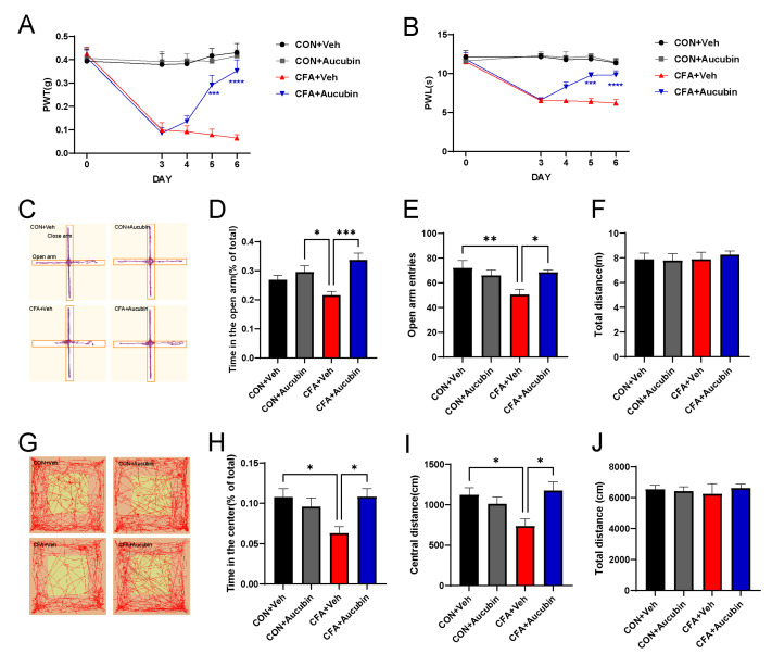
Methods: The CFA-induced inflammatory pain model was employed to assess whether aucubin exerts analgesic effects and its potential mechanisms. Behavioral tests evaluated mechanical and thermal hyperalgesia as well as anxiety-like behaviors in mice. The activation of spinal glial cells and the expression of pro-inflammatory cytokines were examined to evaluate neuroinflammation. Additionally, RNA sequencing was utilized for the identification of differentially expressed genes (DEGs). Molecular biology experiments were conducted to determine the levels of the PINK1 gene and autophagy-related genes, along with PINK1 distribution in neural cells. Furthermore, mitophagy induced by carbonyl cyanide m-chlorophenylhydrazone (CCCP) was employed to examine the roles of PINK1 and mitophagy in pain processing.
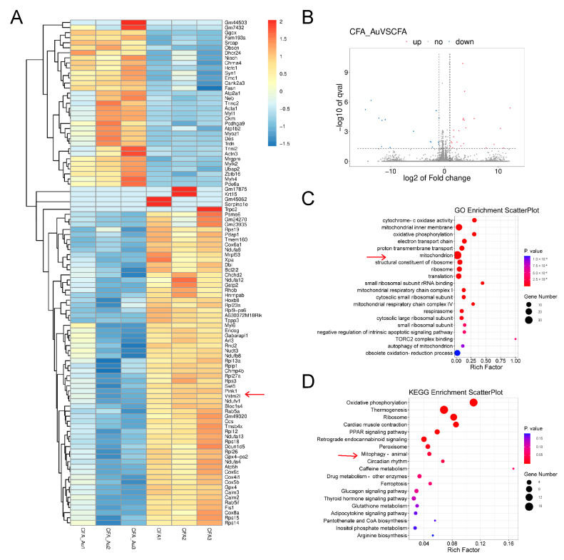
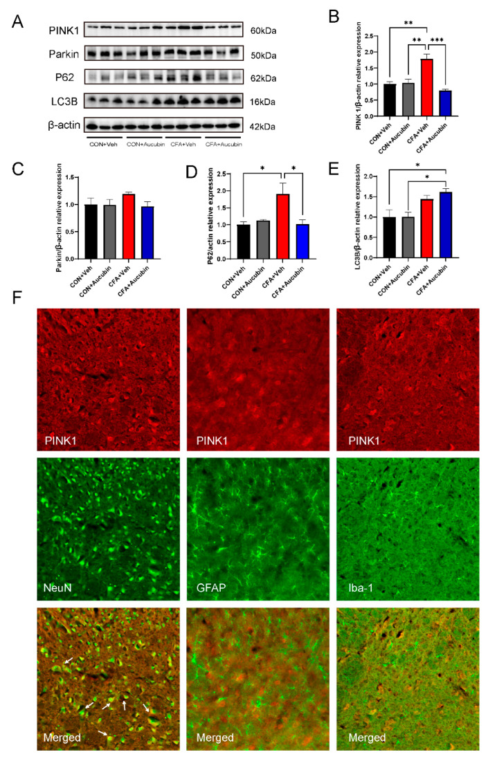
Results: Aucubin significantly ameliorated pain and anxiety-like behaviors induced by CFA in mice and reduced spinal inflammation. RNA sequencing indicated PINK1 as a pivotal gene, and aucubin treatment led to a significant downregulation of PINK1 expression. Further GO and KEGG analyses suggested the involvement of mitochondrial function in the therapeutic regulation of aucubin. Western blotting revealed that aucubin markedly decreased PINK1, Parkin, and p62 levels while increasing LC3B expression. Immunofluorescence showed the predominant co-localization of PINK1 with neuronal cells. Moreover, CCCP-induced mitophagy alleviated mechanical and thermal hyperalgesia caused by CFA and reversed CFA-induced mitochondrial dysfunction.
Conclusions: In summary, our data suggest that aucubin effectively alleviates CFA-induced inflammatory pain, potentially through triggering the PINK1 pathway, promoting mitophagy, and suppressing inflammation. These results provide a novel theoretical foundation for addressing the treatment of inflammatory pain.
Keywords: PINK1; aucubin; inflammation; mitophagy; pain.
Conflict of interest statement
The authors declare no conflict of interest.
Figures


Similar articles
-
[Treadmill exercise alleviates neuropathic pain by regulating mitophagy of the anterior cingulate cortex in rats].Sheng Li Xue Bao. 2023 Apr 25;75(2):160-170. Sheng Li Xue Bao. 2023. PMID: 37089090 Chinese.
-
PGAM5 regulates PINK1/Parkin-mediated mitophagy via DRP1 in CCCP-induced mitochondrial dysfunction.Toxicol Lett. 2018 Mar 1;284:120-128. doi: 10.1016/j.toxlet.2017.12.004. Epub 2017 Dec 11. Toxicol Lett. 2018. PMID: 29241732
-
Tetrahydroxy stilbene glycoside alleviated inflammatory damage by mitophagy via AMPK related PINK1/Parkin signaling pathway.Biochem Pharmacol. 2020 Jul;177:113997. doi: 10.1016/j.bcp.2020.113997. Epub 2020 Apr 27. Biochem Pharmacol. 2020. PMID: 32353422
-
Alleviation of CCCP-induced mitochondrial injury by augmenter of liver regeneration via the PINK1/Parkin pathway-dependent mitophagy.Exp Cell Res. 2021 Dec 1;409(1):112866. doi: 10.1016/j.yexcr.2021.112866. Epub 2021 Oct 13. Exp Cell Res. 2021. PMID: 34655600
-
PINK1/Parkin pathway-mediated mitophagy by AS-IV to explore the molecular mechanism of muscle cell damage.Biomed Pharmacother. 2023 May;161:114533. doi: 10.1016/j.biopha.2023.114533. Epub 2023 Mar 21. Biomed Pharmacother. 2023. PMID: 36948131
Cited by
-
Aucubin mitigates the elevation of microglial aerobic glycolysis and inflammation in diabetic neuropathic pain via aldose reductase.World J Diabetes. 2025 May 15;16(5):103915. doi: 10.4239/wjd.v16.i5.103915. World J Diabetes. 2025. PMID: 40487600 Free PMC article.
-
Microglial metabolic reprogramming: Aucubin inhibits aldose reductase to reverse diabetic neuropathic pain.World J Diabetes. 2025 Aug 15;16(8):110285. doi: 10.4239/wjd.v16.i8.110285. World J Diabetes. 2025. PMID: 40837338 Free PMC article.
References
-
- Djouhri L., Al Otaibi M., Kahlat K., Smith T., Sathish J., Weng X. Persistent hindlimb inflammation induces changes in activation properties of hyperpolarization-activated current (Ih) in rat C-fiber nociceptors in vivo. Neuroscience. 2015;301:121–133. doi: 10.1016/j.neuroscience.2015.05.074. - DOI - PubMed
Grants and funding
LinkOut - more resources
Full Text Sources
