. 2023 Nov 2;16(11):1554.
doi: 10.3390/ph16111554.

Stable Gastric Pentadecapeptide BPC 157 Therapy: Effect on Reperfusion Following Maintained Intra-Abdominal Hypertension (Grade III and IV) in Rats

Affiliations

Stable Gastric Pentadecapeptide BPC 157 Therapy: Effect on Reperfusion Following Maintained Intra-Abdominal Hypertension (Grade III and IV) in Rats

Marijan Tepes et al. Pharmaceuticals (Basel). .

Abstract

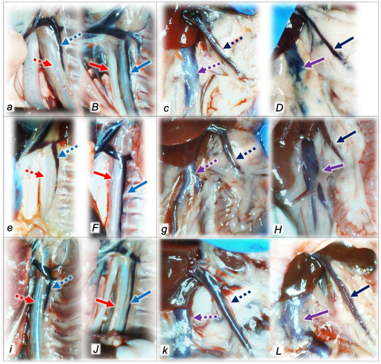
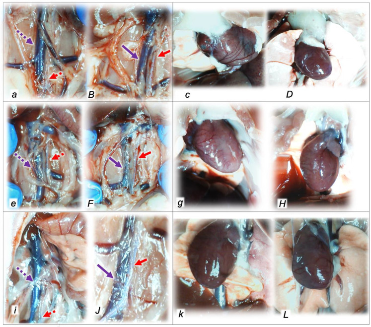
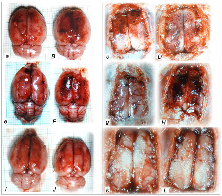
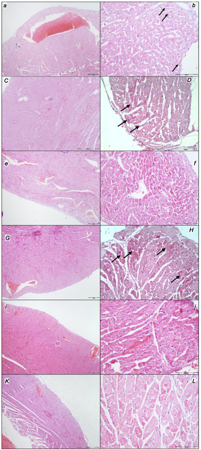
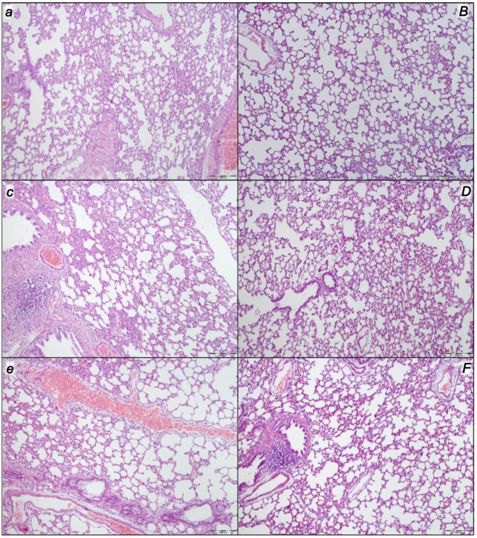
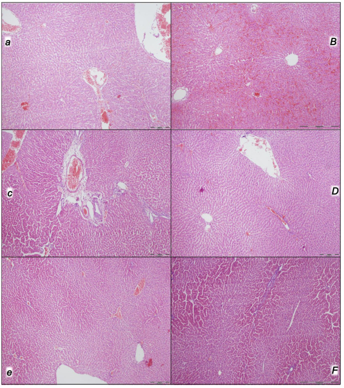
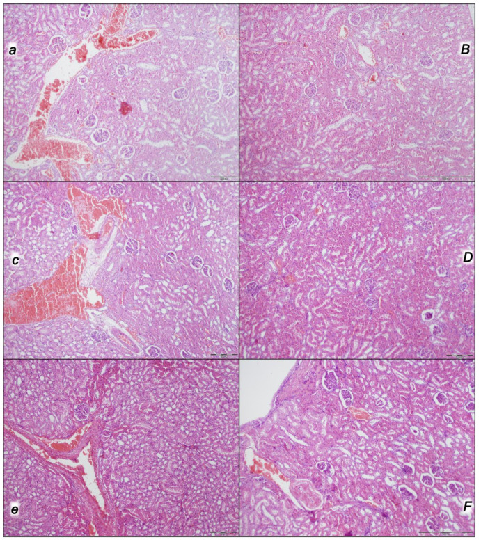
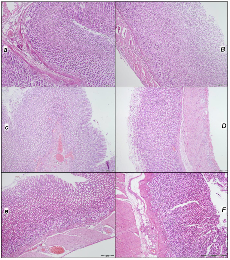
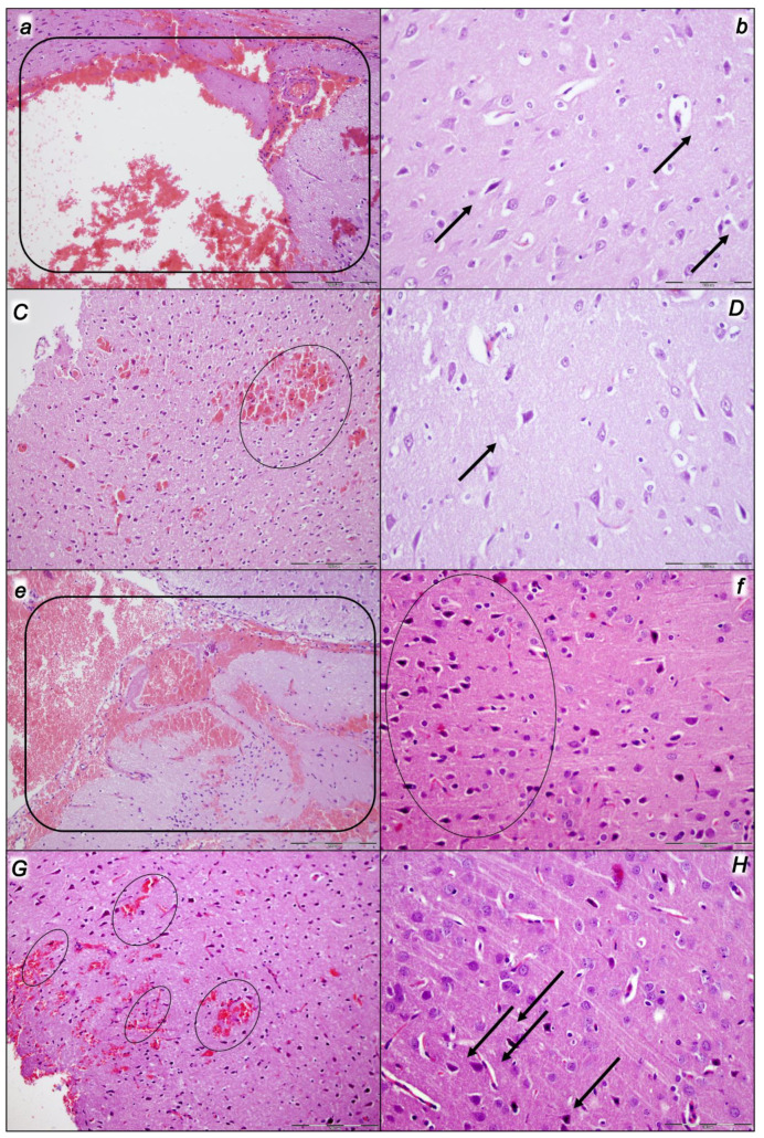
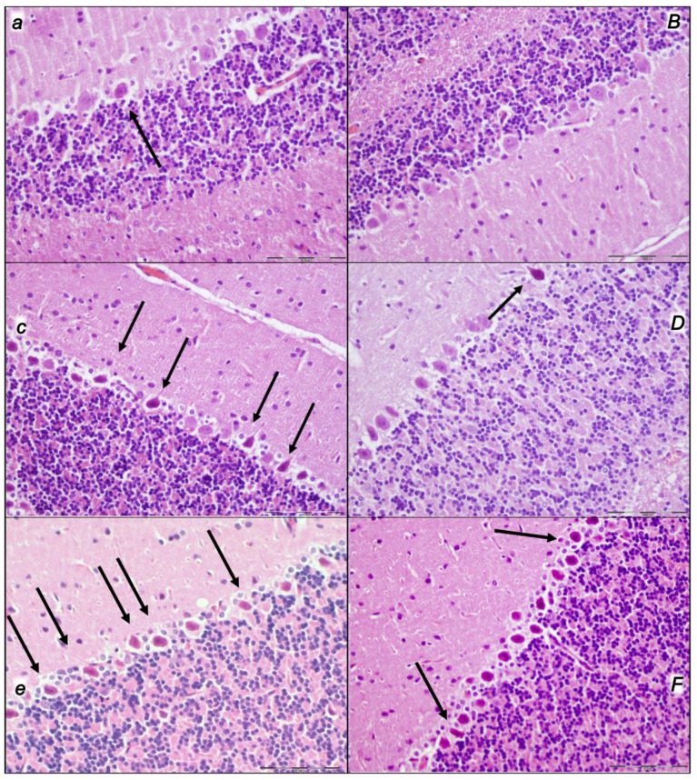
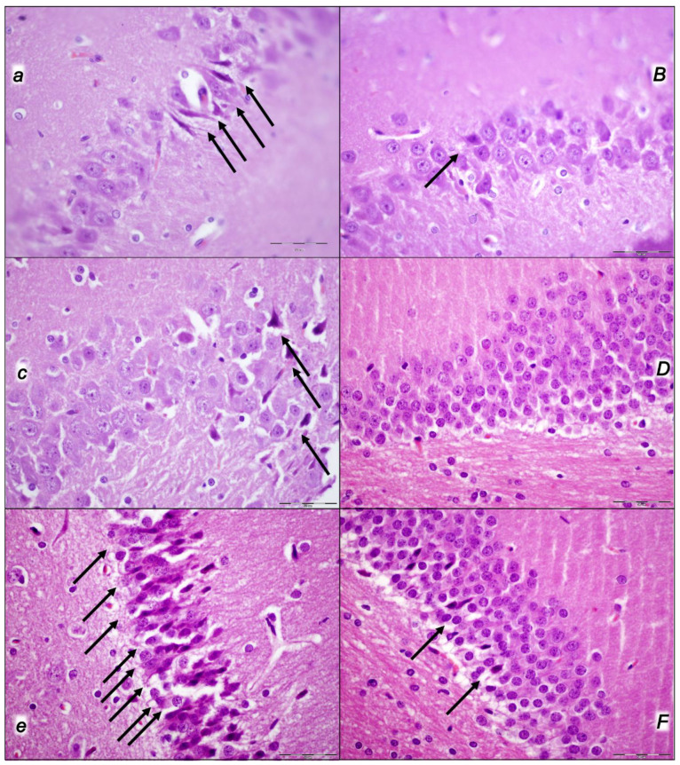
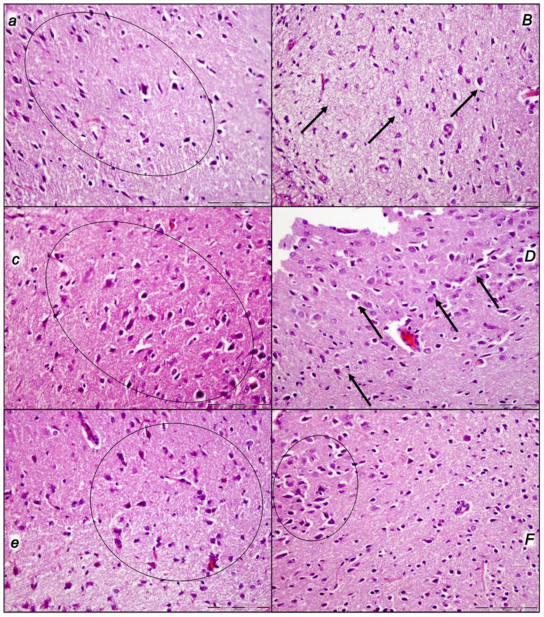
Given in reperfusion, the use of stable gastric pentadecapeptide BPC 157 is an effective therapy in rats. It strongly counteracted, as a whole, decompression/reperfusion-induced occlusion/occlusion-like syndrome following the worst circumstances of acute abdominal compartment and intra-abdominal hypertension, grade III and grade IV, as well as compression/ischemia-occlusion/occlusion-like syndrome. Before decompression (calvariectomy, laparotomy), rats had long-lasting severe intra-abdominal hypertension, grade III (25 mmHg/60 min) (i) and grade IV (30 mmHg/30 min; 40 mmHg/30 min) (ii/iii), and severe occlusion/occlusion-like syndrome. Further worsening was caused by reperfusion for 60 min (i) or 30 min (ii/iii). Severe vascular and multiorgan failure (brain, heart, liver, kidney, and gastrointestinal lesions), widespread thrombosis (peripherally and centrally) severe arrhythmias, intracranial (superior sagittal sinus) hypertension, portal and caval hypertension, and aortal hypotension were aggravated. Contrarily, BPC 157 therapy (10 µg/kg, 10 ng/kg sc) given at 3 min reperfusion times eliminated/attenuated venous hypertension (intracranial (superior sagittal sinus), portal, and caval) and aortal hypotension and counteracted the increases in organ lesions and malondialdehyde values (blood ˃ heart, lungs, liver, kidney ˃ brain, gastrointestinal tract). Vascular recovery promptly occurred (i.e., congested inferior caval and superior mesenteric veins reversed to the normal vessel presentation, the collapsed azygos vein reversed to a fully functioning state, the inferior caval vein-superior caval vein shunt was recovered, and direct blood delivery returned). BPC 157 therapy almost annihilated thrombosis and hemorrhage (i.e., intracerebral hemorrhage) as proof of the counteracted general stasis and Virchow triad circumstances and reorganized blood flow. In conclusion, decompression/reperfusion-induced occlusion/occlusion-like syndrome counteracted by BPC 157 therapy in rats is likely for translation in patients. It is noteworthy that by rapidly counteracting the reperfusion course, it also reverses previous ischemia-course lesions, thus inducing complete recovery.

Keywords: occlusion/occlusion-like syndrome; prime acute abdominal compartment; reperfusion; stable gastric pentadecapeptide BPC 157 therapy.


Conflict of interest statement

The authors declare no conflict of interest.

Figures

Figure 1
Figure 1
Illustrative presentation of blood vessels (a,B,c,D,e,F,g,H,i,J,k,L) in control rats (small italic letters) (a,c,e,g,i,k) and BPC 157-treated rats (capital italic letters) (B,D,F,H,J,L). Considerable failure or recovery was noted after decompression and reperfusion in rats who were subjected to the intra-abdominal hypertension of 25 mmHg for 60 min (a,B,c,D) (i), 30 mmHg for 30 min (e,F,g,H) (ii), or 40 mmHg for 30 min (iii) (i,J,k,L) and sacrificed after the corresponding reperfusion period (60 min (i) (a,B,c,D) or 30 min (ii (e,F,g,H); iii (i,J,k,L))) depending on whether they had received (sc) saline (controls) or BPC 157 at 3 min reperfusion times. Commonly, controls presented the marked congestion of the superior mesenteric vein (dashed black arrows) and inferior caval vein (dashed violet arrows) and a collapsed azygos vein (dashed blue arrows) and aorta (dashed red arrows). Contrarily, BPC 157-treated rats exhibited a consistent therapy effect and counteracted failed vessel presentation; the superior mesenteric vein (full black arrows) and inferior caval vein (full violet arrows) reversed to normal vessel presentation; the collapsed azygos vein (full blue arrows) and aorta (full red arrows) fully recovered.
Figure 2
Figure 2
Illustrative presentation of blood vessels and heart (a,B,c,D,e,F,g,H,i,J,k,L) in control rats (small italic letters) (a,c,e,g,i,k) and BPC 157-treated rats (capital italic letters) (B,D,F,H,J,L). Considerable failure or recovery was noted after decompression and reperfusion in rats who were subjected to the intra-abdominal hypertension of 25 mmHg for 60 min (a,B,c,D) (i), 30 mmHg for 30 min (e,F,g,H) (ii), or 40 mmHg for 30 min (iii) (i,J,k,L) and sacrificed after the corresponding reperfusion period (60 min (i) (a,B,c,D) or 30 min (ii (e,F,g,H); iii (i,J,k,L))) depending on whether they had received (sc) saline (controls) or BPC 157 at 3 min reperfusion times. Commonly, controls presented the marked congestion of the inferior caval vein (dashed violet arrows), a collapsed abdominal aorta (dashed red arrows), and a dilated heart. Contrarily, BPC 157-treated rats exhibited a consistent therapy effect and counteracted failed vessel and heart presentation; the inferior caval vein (full violet arrows) reversed to normal vessel presentation; the abdominal aorta (full red arrows) fully recovered and counteracted heart dilatation.
Figure 3
Figure 3
Illustrative brain presentation, ex vivo and in vivo (a,B,c,D,e,F,g,H,i,J,k,L), in control rats (small italic letters) (a,c,e,g,i,k) and BPC 157-treated rats (capital italic letters) (B,D,F,H,J,L). Considerable gross brain failure (swelling) or recovery (counteracted swelling) was noted after decompression and reperfusion in rats who were subjected to the intra-abdominal hypertension of 25 mmHg for 60 min (a,B,c,D) (i), 30 mmHg for 30 min (e,F,g,H) (ii), or 40 mmHg for 30 min (iii) (i,J,k,L) and sacrificed after the corresponding reperfusion period (60 min (i) (a,B,c,D) or 30 min (ii (e,F,g,H); iii (i,J,k,L))) depending on whether they had received (sc) saline (controls) or BPC 157 at 3 min reperfusion times. Commonly, controls presented marked brain swelling. Contrarily, BPC 157-treated rats exhibited a consistent therapy effect and counteracted brain swelling.
Figure 4
Figure 4
Microscopic changes presented in the heart (a,b,C,D,e,f,G,H,i,j,K,L) in control rats (small italic letters) (a,b,e,f,i,j) and BPC 157-treated rats (capital italic letters) (C,D,G,H,K,L). Considerable lesions were noted after decompression and reperfusion in rats who were subjected to the intra-abdominal hypertension of 25 mmHg for 60 min (a,b,C,D) (i), 30 mmHg for 30 min (e,f,G,H) (ii), or 40 mmHg for 30 min (iii) (i,j,K,L) and sacrificed after the corresponding reperfusion period (60 min (i) (a,b,C,D) or 30 min (ii (e,f,G,H); iii (i,j,K,L)) depending on whether they had received (sc) saline (controls) or BPC 157 at 3 min reperfusion times. Commonly, controls presented the marked congestion of the myocardium with pronounced congestion and the dilatation of coronary arteries and their intramyocardial branches (a,e,i), scattered subendocardial ischemic myocytes (b) (black arrows), and subendocardial infarction (f,j). Contrarily, BPC 157-treated rats exhibited a consistent therapy effect, only mild myocardium congestion (C,G,K), and scattered subendocardial ischemic myocytes (D,H,L) (black arrows). HE staining; magnification, 100×; scale bar, 200 μm (a,C,e,G,i,K) or magnification, 400×; scale bar, 100 μm (b,D,f,H,j,L).
Figure 5
Figure 5
Microscopic changes presented in the lung (a,B,c,D,e,F) in control rats (small italic letters) (a,b,e,f,i,j) and BPC 157-treated rats (capital italic letters) (B,D,F). Considerable lesions were noted after decompression and reperfusion in rats who were subjected to the intra-abdominal hypertension of 25 mmHg for 60 min (a,B) (i), 30 mmHg for 30 min (c,D) (ii), or 40 mmHg for 30 min (iii) (e,F) and sacrificed after the corresponding reperfusion period (60 min (i) (a,B) or 30 min (ii (c,D); iii (e,F)) depending on whether they had received (sc) saline (controls) or BPC 157 at 3 min reperfusion times. Commonly, control animals exhibited the marked congestion of the lung parenchyma, thickening of the alveolar membranes due to capillary congestion, pulmonary edema, and dilatation of larger blood vessels (a,c,e). No changes appeared in the BPC 157-treated rats (B,D,F). HE staining; magnification, 100×; scale bar, 200 μm.
Figure 6
Figure 6
Microscopic changes presented in the liver (a,B,c,D,e,F) in control rats (small italic letters) (a,b,e,f,i,j) and BPC 157-treated rats (capital italic letters) (B,D,F). Considerable lesions were noted after decompression and reperfusion in rats who were subjected to the intra-abdominal hypertension of 25 mmHg for 60 min (a,B) (i), 30 mmHg for 30 min (c,D) (ii), or 40 mmHg for 30 min (iii) (e,F) and sacrificed after the corresponding reperfusion period (60 min (i) (a,B) or 30 min (ii (c,D); iii (e,F)) upon receiving (sc) saline (controls) or BPC 157 at 3 min reperfusion times. Commonly, control animals exhibited the marked congestion of the liver parenchyma, with a pronounced dilatation of sinusoids and branches of the portal vein in portal tracts (a,c,e). No changes or mild congestion appeared in the BPC 157-treated rats (B,D,F). HE staining; magnification, 100×; scale bar, 200 μm.
Figure 7
Figure 7
Microscopic changes presented in the kidney (a,B,c,D,e,F) in control rats (small italic letters) (a,b,e,f,i,j) and BPC 157-treated rats (capital italic letters) (B,D,F). Considerable lesions were noted after decompression and reperfusion in rats who were subjected to the intra-abdominal hypertension of 25 mmHg for 60 min (a,B) (i), 30 mmHg for 30 min (c,D) (ii), or 40 mmHg for 30 min (iii) (e,F) and sacrificed after the corresponding reperfusion period (60 min (i) (a,B) or 30 min (ii (c,D); iii (e,F)) upon receiving (sc) saline (controls) or BPC 157 at 3 min reperfusion times. Commonly, control animals exhibited the marked congestion of the renal parenchyma with moderate vascular congestion, and interstitial edema occurred in control rats (a,c,e). No changes or mild congestion appeared in the BPC 157-treated rats (B,D,F). HE staining; magnification, 100×; scale bar, 200 μm.
Figure 8
Figure 8
Microscopic changes presented in the stomach (a,B,c,D,e,F) in control rats (small italic letters) (a,b,e,f,i,j) and BPC 157-treated rats (capital italic letters) (B,D,F). Considerable lesions were noted after decompression and reperfusion in rats who were subjected to the intra-abdominal hypertension of 25 mmHg for 60 min (a,B) (i), 30 mmHg for 30 min (c,D) (ii), or 40 mmHg for 30 min (iii) (e,F) and sacrificed after the corresponding reperfusion period (60 min (i) (a, B) or 30 min (ii (c,D); iii (e,F)) depending on whether they had received (sc) saline (controls) or BPC 157 at 3 min reperfusion times. Commonly, control animals exhibited the marked congestion of the stomach wall due to the transmural pronounced congestion and dilatation of the blood vessels (a,c,e). No changes appeared in the BPC 157-treated rats (B,D,F). HE staining; magnification, 100×; scale bar, 200 μm.
Figure 9
Figure 9
Neuropathological changes presented in the cerebrum (a,b,C,D,e,f,G,H) in control rats (small italic letters) (a,b,e,f) and BPC 157-treated rats (capital italic letters) (C,D,G,H). Considerable lesions were noted after decompression and reperfusion in rats who were subjected to the intra-abdominal hypertension of 25 mmHg for 60 min (a,b,C,D) (i) or 40 mmHg for 30 min (ii) (e,f,G,H) and sacrificed after the corresponding reperfusion period (60 min (i) (a,b,C,D) or 30 min (ii)) (e,f,G,H) depending on whether they had received (sc) saline (controls) or BPC 157 at 3 min reperfusion times. Commonly, controls presented a pronounced edema and congestion in the brain tissue. In the BPC 157-treated rats (capital italic letters), only mild edema in the brain tissue was found. A focal and deeper neocortical hemorrhage was found in control animals affecting the neocortex, corpus callosum, amygdala, and striatum in the brain tissue (marked areas—(i): (a); (ii): (e)). In the BPC 157 group, only smaller areas of neocortical hemorrhage occurred (marked areas—(i): (C); (ii): (G)). Moderate to severe neurodegenerative changes were presented in the cerebral cortex, along with the karyopyknosis of cortical neurons, in controls (marked areas, black arrows) ((i): (b); (ii): (f)) while BPC 157 rats exhibited, consistently, only rare karyopyknotic cells and mild neurodegenerative changes ((i): (D); (ii): (H)). HE staining; magnification, 200×; scale bar, 200 μm.
Figure 10
Figure 10
Neuropathological changes presented in the cerebellum (a,B,c,D,e,F) in control rats (small italic letters) (a,c,e) and BPC 157-treated rats (capital italic letters) (B,D,F). Considerable lesions were noted after decompression and reperfusion in rats who were subjected to the intra-abdominal hypertension of 25 mmHg for 60 min (a,B) (i), 30 mmHg for 30 min (ii) (c,D), or 40 mmHg for 30 min (iii) (e,F) and sacrificed after the corresponding reperfusion period (60 min (i) (a,B) or 30 min ((ii) (c,D), and (iii) (e,F))) depending on whether they had received (sc) saline (controls) or BPC 157 at 3 min reperfusion times. Commonly, in the brain tissue, pronounced edema and congestion occurred in controls. Moderate neurodegenerative changes in the cerebellar cortex were found in control animals. There were karyopyknosis and the degeneration of Purkinje cells of the cerebellar cortex (black arrows). In BPC 157 rats, only mild edema in the brain tissue was found, and only rare karyopyknotic cells and mild neurodegenerative changes in the cerebellar cortex occurred (black arrows). HE staining; magnification, 400×; scale bar, 100 μm.
Figure 11
Figure 11
Neuropathological changes presented in the hippocampus (a,B,c,D,e,F) in control rats (small italic letters) (a,c,e) and BPC 157-treated rats (capital italic letters) (B,D,F). Considerable lesions were noted after decompression and reperfusion in rats who were subjected to the intra-abdominal hypertension of 25 mmHg for 60 min (a,B) (i), 30 mmHg for 30 min (ii) (c,D), or 40 mmHg for 30 min (iii) (e,F) and sacrificed after the corresponding reperfusion period (60 min (i) (a,B), 30 min ((ii) (c,D) and (iii) (e,F))) depending on whether they had received (sc) saline (controls) or BPC 157 at 3 min reperfusion times. Commonly, in the brain tissue, pronounced edema and congestion occurred in controls. In the hippocampus, there were moderate neurodegenerative changes in the control animals. There were karyopyknosis and the degeneration of pyramidal cells of the hippocampus (black arrows). BPC 157 rats exhibited only mild edema in the brain tissue and no or only rare karyopyknotic cells in the hippocampus (black arrows). HE staining; magnification, 600×, scale bar, 50 μm.
Figure 12
Figure 12
Neuropathological changes presented in the hypothalamus (a,B,c,D,e,F) in control rats (small italic letters) (a,c,e) and BPC 157-treated rats (capital italic letters) (B,D,F). Considerable lesions were noted after decompression and reperfusion in rats who were subjected to the intra-abdominal hypertension of 25 mmHg for 60 min (a,B) (i), 30 mmHg for 30 min (ii) (c,D), or 40 mmHg for 30 min (iii) (e,F) and sacrificed after the corresponding reperfusion period (60 min (i) (a,B), 30 min ((ii) (c,D) and (iii) (e,F))) depending on whether they had received (sc) saline (controls) or BPC 157 at 3 min reperfusion times. Commonly, in the brain tissue, pronounced edema and congestion occurred in controls. In the hippocampus, there were moderate to severe neurodegenerative changes in the control animals. There were karyopyknosis hypothalamic neurons (black arrows). BPC 157 rats exhibited only mild edema in the brain tissue and only rare karyopyknotic cells and mild neurodegenerative changes in the hypothalamus (black arrows). HE staining; magnification, 400×; scale bar, 100 μm.

Similar articles

Cited by

References

    1. Sikiric P., Skrtic A., Gojkovic S., Krezic I., Zizek H., Lovric E., Sikiric S., Knezevic M., Strbe S., Milavic M., et al. Cytoprotective gastric pentadecapeptide BPC 157 resolves major vessel occlusion disturbances, ischemia-reperfusion injury following Pringle maneuver, and Budd-Chiari syndrome. World J. Gastroenterol. 2022;28:23–46. doi: 10.3748/wjg.v28.i1.23. - DOI - PMC - PubMed
    1. Sikiric P., Hahm K.B., Blagaic A.B., Tvrdeic A., Pavlov K.H., Petrovic A., Kokot A., Gojkovic S., Krezic I., Drmic D., et al. Stable gastric pentadecapeptide BPC 157, Robert’s stomach cytoprotection/adaptive cytoprotection/organoprotection, and Selye’s stress coping response: Progress, achievements, and the future. Gut Liver. 2020;14:153–167. doi: 10.5009/gnl18490. - DOI - PMC - PubMed
    1. Sikiric P., Rucman R., Turkovic B., Sever M., Klicek R., Radic B., Drmic D., Stupnisek M., Misic M., Vuletic L.B., et al. Novel cytoprotective mediator, stable gastric pentadecapeptide BPC 157. Vascular recruitment and gastrointestinal tract healing. Curr. Pharm. Des. 2018;24:1990–2001. doi: 10.2174/1381612824666180608101119. - DOI - PubMed
    1. Vukojevic J., Milavic M., Perovic D., Ilic S., Zemba Cilic A., Duran N., Strbe S., Zoricic Z., Filipcic I., Brecic P., et al. Pentadecapeptide BPC 157 and the central nervous system. Neural Regen. Res. 2022;17:482–487. doi: 10.4103/1673-5374.320969. - DOI - PMC - PubMed
    1. Seiwerth S., Milavic M., Vukojevic J., Gojkovic S., Krezic I., Vuletic L.B., Pavlov K.H., Petrovic A., Sikiric S., Vranes H., et al. Stable gastric pentadecapeptide BPC 157 and wound healing. Front. Pharmacol. 2021;12:627533. doi: 10.3389/fphar.2021.627533. - DOI - PMC - PubMed

LinkOut - more resources