

Synthesis of Pyrrolo[3,4- b]pyridin-5-ones via Ugi-Zhu Reaction and In Vitro-In Silico Studies against Breast Carcinoma
- PMID: 38004428
- PMCID: PMC10674953
- DOI: 10.3390/ph16111562
Synthesis of Pyrrolo[3,4- b]pyridin-5-ones via Ugi-Zhu Reaction and In Vitro-In Silico Studies against Breast Carcinoma
Abstract
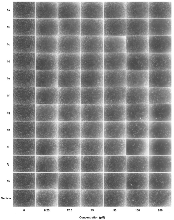
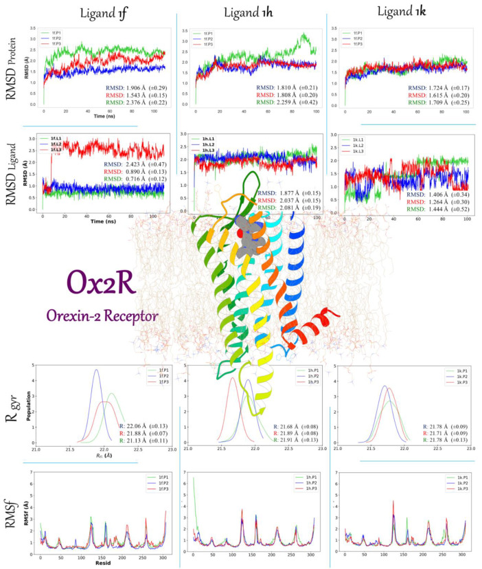
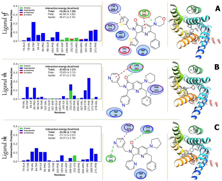
An Ugi-Zhu three-component reaction (UZ-3CR) coupled in a one-pot manner to a cascade process (N-acylation/aza Diels-Alder cycloaddition/decarboxylation/dehydration) was performed to synthesize a series of pyrrolo[3,4-b]pyridin-5-ones in 20% to 92% overall yields using ytterbium triflate as a catalyst, toluene as a solvent, and microwaves as a heat source. The synthesized molecules were evaluated in vitro against breast cancer cell lines MDA-MB-231 and MCF-7, finding that compound 1f, at a concentration of 6.25 μM, exhibited a potential cytotoxic effect. Then, to understand the interactions between synthesized compounds and the main proteins related to the cancer cell lines, docking studies were performed on the serine/threonine kinase 1 (AKT1) and Orexetine type 2 receptor (Ox2R), finding moderate to strong binding energies, which matched accurately with the in vitro results. Additionally, molecular dynamics were performed between proteins related to the studied cell lines and the three best ligands.
Keywords: MCF-7; MCRs; MDA-MB-231; Pyrrolo[3,4-b]pyridin-5-ones; Ugi–Zhu; breast cancer; docking; molecular dynamics.
Conflict of interest statement
The authors declare no conflict of interest. The funders had no role in the design of the study; in the collection, analyses, or interpretation of data; in the writing of the manuscript; or in the decision to publish the results.
Figures


Similar articles
-
Multi-Component Synthesis of New Fluorinated-Pyrrolo[3,4-b]pyridin-5-ones Containing the 4-Amino-7-chloroquinoline Moiety and In Vitro-In Silico Studies Against Human SARS-CoV-2.Int J Mol Sci. 2025 Aug 7;26(15):7651. doi: 10.3390/ijms26157651. Int J Mol Sci. 2025. PMID: 40806777 Free PMC article.
-
Synthesis of bis-furyl-pyrrolo[3,4-b]pyridin-5-ones via Ugi-Zhu reaction and in vitro activity assays against human SARS-CoV-2 and in silico studies on its main proteins.RSC Med Chem. 2022 Nov 18;14(1):154-165. doi: 10.1039/d2md00350c. eCollection 2023 Jan 25. RSC Med Chem. 2022. PMID: 36760742 Free PMC article.
-
Synthesis of New Polyheterocyclic Pyrrolo[3,4-b]pyridin-5-ones via an Ugi-Zhu/Cascade/Click Strategy.Molecules. 2023 May 14;28(10):4087. doi: 10.3390/molecules28104087. Molecules. 2023. PMID: 37241828 Free PMC article.
-
Synthesis of Polyheterocyclic Pyrrolo[3,4-b]pyridin-5-ones via a One-Pot (Ugi-3CR/aza Diels-Alder/N-acylation/aromatization/SN2) Process. A Suitable Alternative towards Novel Aza-Analogues of Falipamil.Molecules. 2018 Mar 27;23(4):763. doi: 10.3390/molecules23040763. Molecules. 2018. PMID: 29584639 Free PMC article.
-
In-vitro Evaluation of Isatin Derivatives as Potent Anti-Breast Cancer Agents against MCF-7, MDA MB 231, MDA-MB 435 and MDA-MB 468 Breast Cancers Cell Lines: A Review.Anticancer Agents Med Chem. 2022;22(10):1883-1896. doi: 10.2174/1871520621666210903130152. Anticancer Agents Med Chem. 2022. PMID: 34477529 Review.
Cited by
-
Multi-Component Synthesis of New Fluorinated-Pyrrolo[3,4-b]pyridin-5-ones Containing the 4-Amino-7-chloroquinoline Moiety and In Vitro-In Silico Studies Against Human SARS-CoV-2.Int J Mol Sci. 2025 Aug 7;26(15):7651. doi: 10.3390/ijms26157651. Int J Mol Sci. 2025. PMID: 40806777 Free PMC article.
References
Grants and funding
LinkOut - more resources
Full Text Sources
Miscellaneous
