. 2023 Oct 27;15(11):2539.
doi: 10.3390/pharmaceutics15112539.

Selegiline Modulates Lipid Metabolism by Activating AMPK Pathways of Epididymal White Adipose Tissues in HFD-Fed Obese Mice

Affiliations

Selegiline Modulates Lipid Metabolism by Activating AMPK Pathways of Epididymal White Adipose Tissues in HFD-Fed Obese Mice

Hye-Young Joung et al. Pharmaceutics. .

Abstract

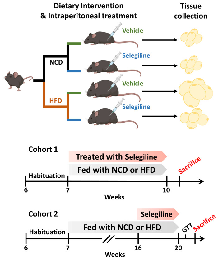
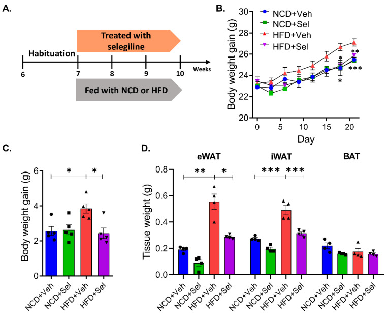
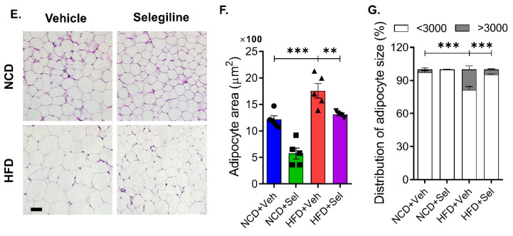
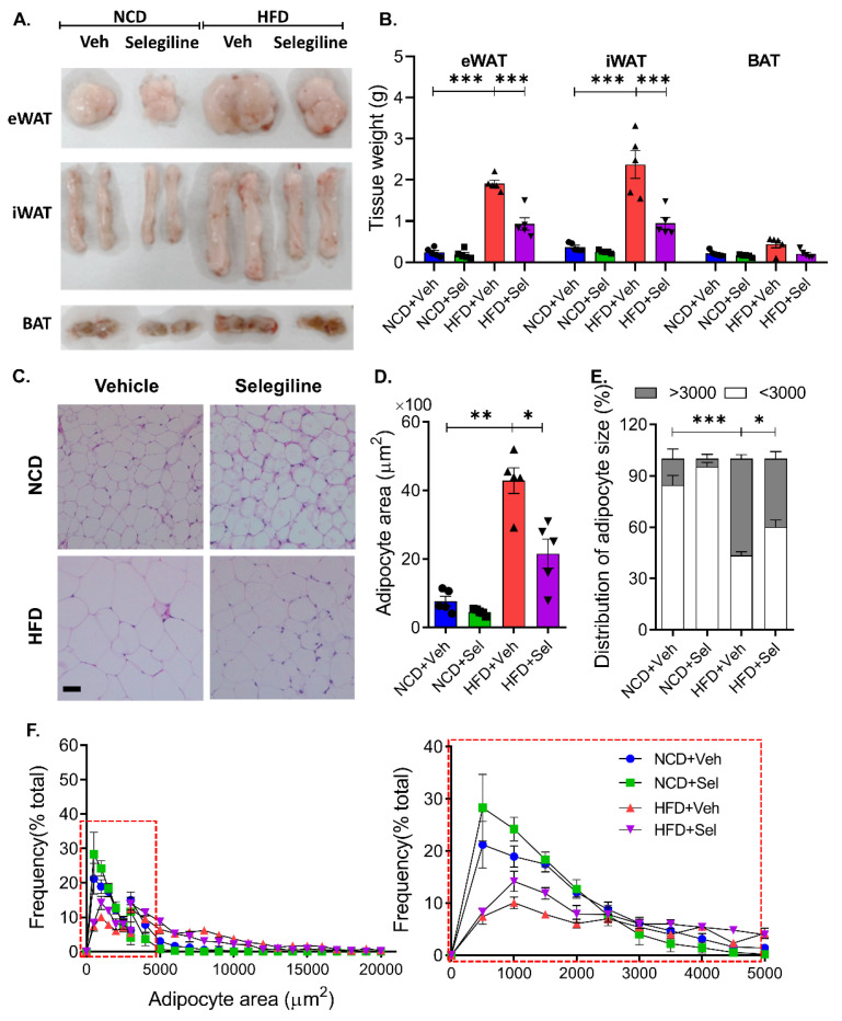
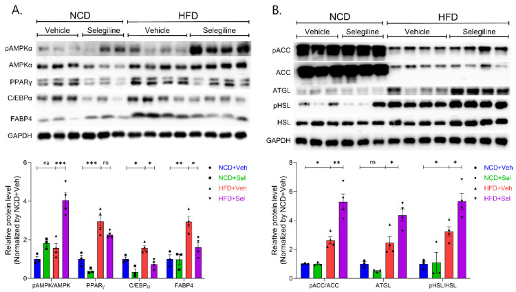
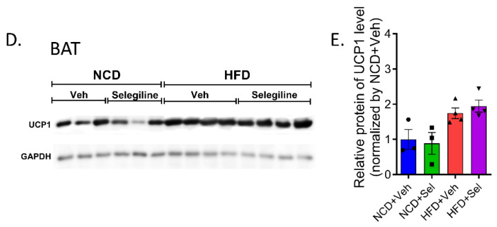
Obesity, as a major cause of many chronic diseases such as diabetes, cardiovascular disease, and cancer, is among the most serious health problems. Increased monoamine oxidase (MAO) activity has been observed in the adipose tissue of obese humans and animals. Although previous studies have already demonstrated the potential of MAO-B inhibitors as a treatment for this condition, the mechanism of their effect has been insufficiently elucidated. In this study, we investigated the anti-obesity effect of selegiline, a selective MAO-B inhibitor, using in vivo animal models. The effect was evaluated through an assessment of body energy homeostasis, glucose tolerance tests, and biochemical analysis. Pharmacological inhibition of MAO-B by selegiline was observed to reduce body weight and fat accumulation, and improved glucose metabolism without a corresponding change in food intake, in HFD-fed obese mice. We also observed that both the expression of adipogenenic markers, including C/EBPα and FABP4, and lipogenic markers such as pACC were significantly reduced in epididymal white adipose tissues (eWATs). Conversely, increased expression of lipolytic markers such as ATGL and pHSL and AMPK phosphorylation were noted. Treating obese mice with selegiline significantly increased expression levels of UCP1 and promoted eWAT browning, indicating increased energy expenditure. These results suggest that selegiline, by inhibiting MAO-B activity, is a potential anti-obesity treatment.

Keywords: adipogenesis; anti-obesity; eWAT browning; lipogenesis; lipolysis; selegiline.


Conflict of interest statement

The authors declare no conflict of interest.

Figures

Figure 1
Figure 1
Schematic illustration of the experimental configuration.
Figure 2
Figure 2
Selegiline prevented weight gain in HFD-fed mice. (A) Schematic diagram for this experiment. C57BL/6 mice were fed NCD or HFD and intraperitoneally treated with vehicle or selegiline (30 mg/kg) once a day for 3 weeks. (B) Body weight curves of vehicle- and selegiline-treated mice fed NCD or HFD (n = 5 per group). (C) Graph of body weight gain each groups. (D) Bar graph of weight of eWATs, iWATs, and BATs. (E) Representative of H&E staining of eWATs. Scale bar = 50 μm. (F) Summary plot of average of adipocyte areas. (G) Summary plot of frequency of large adipocytes, defined as having an adipocyte area greater than 3000 μm2. Data are presented as the mean ± SEM and one-way ANOVA followed by Dunnett’s multiple comparison test was employed for data analysis. Significance denoted by *: p < 0.05, **: p < 0.01, ***: p < 0.001 compared to HFD+Veh. NCD, normal chow diet; HFD, high-fat diet; Veh, vehicle; Sel, selegiline; eWAT, epididymal white adipose tissues; iWAT, inguinal white adipose tissue; BAT, brown adipose tissue.
Figure 2
Figure 2
Selegiline prevented weight gain in HFD-fed mice. (A) Schematic diagram for this experiment. C57BL/6 mice were fed NCD or HFD and intraperitoneally treated with vehicle or selegiline (30 mg/kg) once a day for 3 weeks. (B) Body weight curves of vehicle- and selegiline-treated mice fed NCD or HFD (n = 5 per group). (C) Graph of body weight gain each groups. (D) Bar graph of weight of eWATs, iWATs, and BATs. (E) Representative of H&E staining of eWATs. Scale bar = 50 μm. (F) Summary plot of average of adipocyte areas. (G) Summary plot of frequency of large adipocytes, defined as having an adipocyte area greater than 3000 μm2. Data are presented as the mean ± SEM and one-way ANOVA followed by Dunnett’s multiple comparison test was employed for data analysis. Significance denoted by *: p < 0.05, **: p < 0.01, ***: p < 0.001 compared to HFD+Veh. NCD, normal chow diet; HFD, high-fat diet; Veh, vehicle; Sel, selegiline; eWAT, epididymal white adipose tissues; iWAT, inguinal white adipose tissue; BAT, brown adipose tissue.
Figure 3
Figure 3
Selegiline markedly reduced body weight and fat accumulation in HFD-fed obese mice. (A) Schematic diagram for this experiment. C57BL/6 mice were fed an NCD or HFD for 9 weeks, and then intraperitoneal administrated 30 mg/kg of selegiline or saline once a day for 4 weeks. (B) Representative figure of body weight change in each group. (C) Polled data of body weight of each group. One-way ANOVA followed by Dunnett’s multiple comparison test. (D) Graph of body weight gain by each group. One-way ANOVA followed by Dunnett’s multiple comparison test. (E) Food intake of each group. (F) Left: Polled data of GTT of each group. Two-way RM ANOVA followed by Dunnett’s multiple comparison test, interaction F(6,108) = 5.398, p < 0.0001, time F(6,108) = 351.8, p < 0.0001, between group F(3,18) = 71.06, p < 0.0001. Right: Bar graphs showing area under the curve (AUC) values obtained from GTT experiments. Data are presented as the mean ± SEM. Significance was denoted by *: p < 0.05, **: p < 0.01, ***: p < 0.001 compared to HFD+Veh. NCD, normal chow diet; HFD, high-fat diet; Veh, vehicle; Sel, selegiline.
Figure 4
Figure 4
Selegiline administration reduced epididymal adipocyte size in HFD-fed obese mice. (A) Representative figure of eWATs, iWATs, and BATs. (B) Polled data of weight of eWATs, iWATs, and BATs. (C) Representative images of H&E staining of eWATs from each group. Scale bar = 50 μm. (D) Average of adipocyte size of WAT were measured from H&E images using AdipoArea software. n = 5 per group. (E) Frequency of large adipocytes, defined as having an adipocyte area greater than 3000 μm2. (F) Summary plot of quantitative analysis of adipocyte area. Data are presented as the mean ± SEM and one-way ANOVA followed by Dunnett’s multiple comparison test was employed for data analysis. Significance denoted by *: p < 0.05, **: p < 0.01, ***: p < 0.001 compared to HFD+Veh. NCD, normal chow diet; HFD, high fat diet; Veh (V), vehicle; Sel, selegiline; eWAT, epididymal white adipose tissues; iWAT, inguinal white adipose tissue; BAT, brown adipose tissue.
Figure 5
Figure 5
Selegiline-regulated lipid metabolism in epididymal WATs. (A) Upper panel: Images of Western blot of pAMPK, AMPK, PPARγ, C/EBPα, FABP4, and GAPDH in WATs from mice treated with selegiline or vehicle fed an NCD or HFD. Lower panel: Summary plot of expression of ratio of pAMPK/AMPK, PPARγ, C/EBPα, and FABP4. (B) Upper panel: Images of Western blot of pACC, ACC, ATGL, pHSL, HSL, and GAPDH in eWATs from mice treated with selegiline or vehicle fed an NCD or HFD. Lower panel: Summary plot of expression of ration of pACC/ACC, ATGL, ratio of pHSL/HSL. Data are presented as the mean ± SEM and one-way ANOVA followed by by Dunnett’s multiple comparison test was employed for data analysis. Significance denoted by *: p < 0.05, **: p < 0.01, ***: p < 0.001 compared to HFD+Veh. p(Phospho)AMPKα, AMP-activated protein kinase α; PPARγ, Peroxisome proliferator activated receptor gamma; C/EBPα, CCAAT/enhancer-binding protein alpha; FABP4, fatty acid binding protein 4; GAPDH, Glyceraldehyde-3-phosphate dehydrogenase; pACC, Phospho-Acetyl-CoA Carboxylase; ATGL, Adipose triglyceride lipase; HSL, Hormone-sensitive lipase.
Figure 6
Figure 6
Selegiline promoted thermogenesis and epididymal WAT browning in HFD-fed obese mice. (A) Images of Western blot of PRDM16, PGC1α, UCP1, and GAPDH in eWAT from mice treated with selegiline or vehicle fed an NCD or HFD. (B) Summary plot of expression of PRDM16, PGC1α, and UCP1 in eWATs from mice treated with selegiline or vehicle fed an NCD or HFD. (C) Representative of UCP1 immunostaining of eWAT. Scale bar = 50 μm. (D) Images of Western blot UCP1 and GAPDH in BATs from mice treated with selegiline or vehicle fed an NCD or HFD. (E) Summary plot of expression UCP1 in BATs from mice treated with selegiline or vehicle fed an NCD or HFD. Data are presented as the mean ± SEM and one-way ANOVA followed by Dunnett’s multiple comparison test was employed for data analysis. Significance denoted by *: p < 0.05, **: p < 0.01, ***: p < 0.001 compared to HFD+Veh. Prdm16, PR/SET Domain 16; PGC1α, Peroxisome proliferator-activated receptor gamma coactivator 1-alpha; UCP1, Uncoupling Protein 1; GAPDH, Glyceraldehyde-3-phosphate dehydrogenase; NCD, normal chow diet; HFD, high-fat diet; Veh, vehicle; Sel, selegiline; BAT, brown adipose tissue.
Figure 6
Figure 6
Selegiline promoted thermogenesis and epididymal WAT browning in HFD-fed obese mice. (A) Images of Western blot of PRDM16, PGC1α, UCP1, and GAPDH in eWAT from mice treated with selegiline or vehicle fed an NCD or HFD. (B) Summary plot of expression of PRDM16, PGC1α, and UCP1 in eWATs from mice treated with selegiline or vehicle fed an NCD or HFD. (C) Representative of UCP1 immunostaining of eWAT. Scale bar = 50 μm. (D) Images of Western blot UCP1 and GAPDH in BATs from mice treated with selegiline or vehicle fed an NCD or HFD. (E) Summary plot of expression UCP1 in BATs from mice treated with selegiline or vehicle fed an NCD or HFD. Data are presented as the mean ± SEM and one-way ANOVA followed by Dunnett’s multiple comparison test was employed for data analysis. Significance denoted by *: p < 0.05, **: p < 0.01, ***: p < 0.001 compared to HFD+Veh. Prdm16, PR/SET Domain 16; PGC1α, Peroxisome proliferator-activated receptor gamma coactivator 1-alpha; UCP1, Uncoupling Protein 1; GAPDH, Glyceraldehyde-3-phosphate dehydrogenase; NCD, normal chow diet; HFD, high-fat diet; Veh, vehicle; Sel, selegiline; BAT, brown adipose tissue.

Similar articles

Cited by

References

    1. Jung D.Y., Suh N., Jung M.H. Tanshinone 1 prevents high fat diet-induced obesity through activation of brown adipocytes and induction of browning in white adipocytes. Life Sci. 2022;298:120488. doi: 10.1016/j.lfs.2022.120488. - DOI - PubMed
    1. Khafagy R., Dash S. Obesity and Cardiovascular Disease: The Emerging Role of Inflammation. Front. Cardiovasc. Med. 2021;8:768119. doi: 10.3389/fcvm.2021.768119. - DOI - PMC - PubMed
    1. Zhang F., Ai W., Hu X., Meng Y., Yuan C., Su H., Wang L., Zhu X., Gao P., Shu G., et al. Phytol stimulates the browning of white adipocytes through the activation of AMP-activated protein kinase (AMPK) alpha in mice fed high-fat diet. Food Funct. 2018;9:2043–2050. doi: 10.1039/C7FO01817G. - DOI - PubMed
    1. Shin S.K., Cho H.W., Song S.E., Im S.S., Bae J.H., Song D.K. Oxidative stress resulting from the removal of endogenous catalase induces obesity by promoting hyperplasia and hypertrophy of white adipocytes. Redox Biol. 2020;37:101749. doi: 10.1016/j.redox.2020.101749. - DOI - PMC - PubMed
    1. Ahmad B., Serpell C.J., Fong I.L., Wong E.H. Molecular Mechanisms of Adipogenesis: The Anti-adipogenic Role of AMP-Activated Protein Kinase. Front. Mol. Biosci. 2020;7:76. doi: 10.3389/fmolb.2020.00076. - DOI - PMC - PubMed