Review
. 2023 Nov 2;15(11):2574.
doi: 10.3390/pharmaceutics15112574.

Exploiting Benefits of Vaterite Metastability to Design Degradable Systems for Biomedical Applications

Affiliations
Review

Exploiting Benefits of Vaterite Metastability to Design Degradable Systems for Biomedical Applications

Yulia Svenskaya et al. Pharmaceutics. .

Abstract

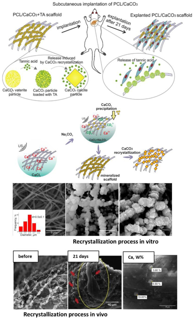
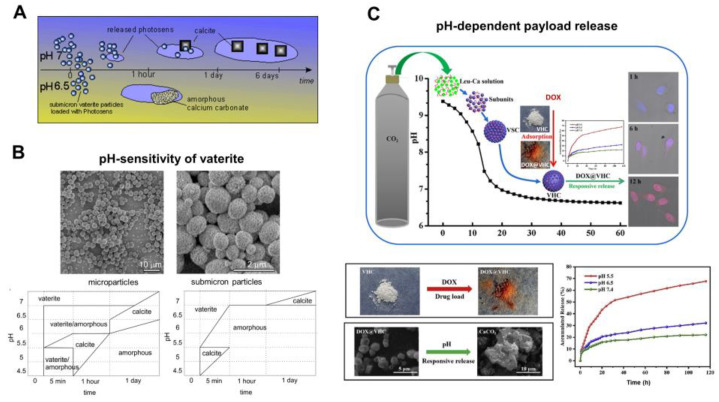
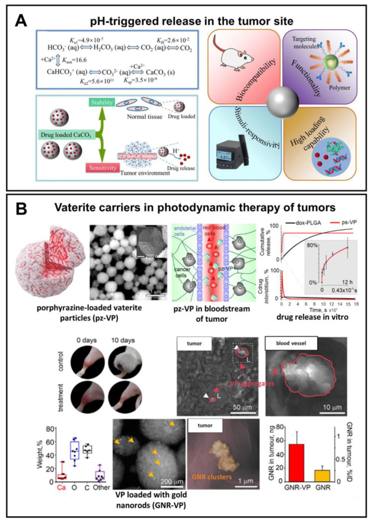
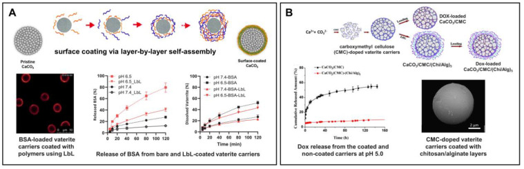
The widespread application of calcium carbonate is determined by its high availability in nature and simplicity of synthesis in laboratory conditions. Moreover, calcium carbonate possesses highly attractive physicochemical properties that make it suitable for a wide range of biomedical applications. This review provides a conclusive analysis of the results on using the tunable vaterite metastability in the development of biodegradable drug delivery systems and therapeutic vehicles with a controlled and sustained release of the incorporated cargo. This manuscript highlights the nuances of vaterite recrystallization to non-porous calcite, dissolution at acidic pH, biodegradation at in vivo conditions and control over these processes. This review outlines the main benefits of vaterite instability for the controlled liberation of the encapsulated molecules for the development of biodegradable natural and synthetic polymeric materials for biomedical purposes.

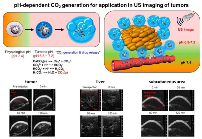
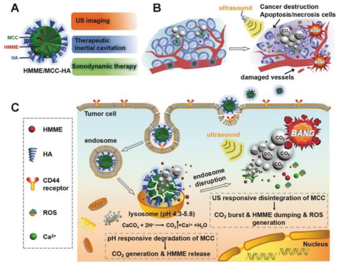
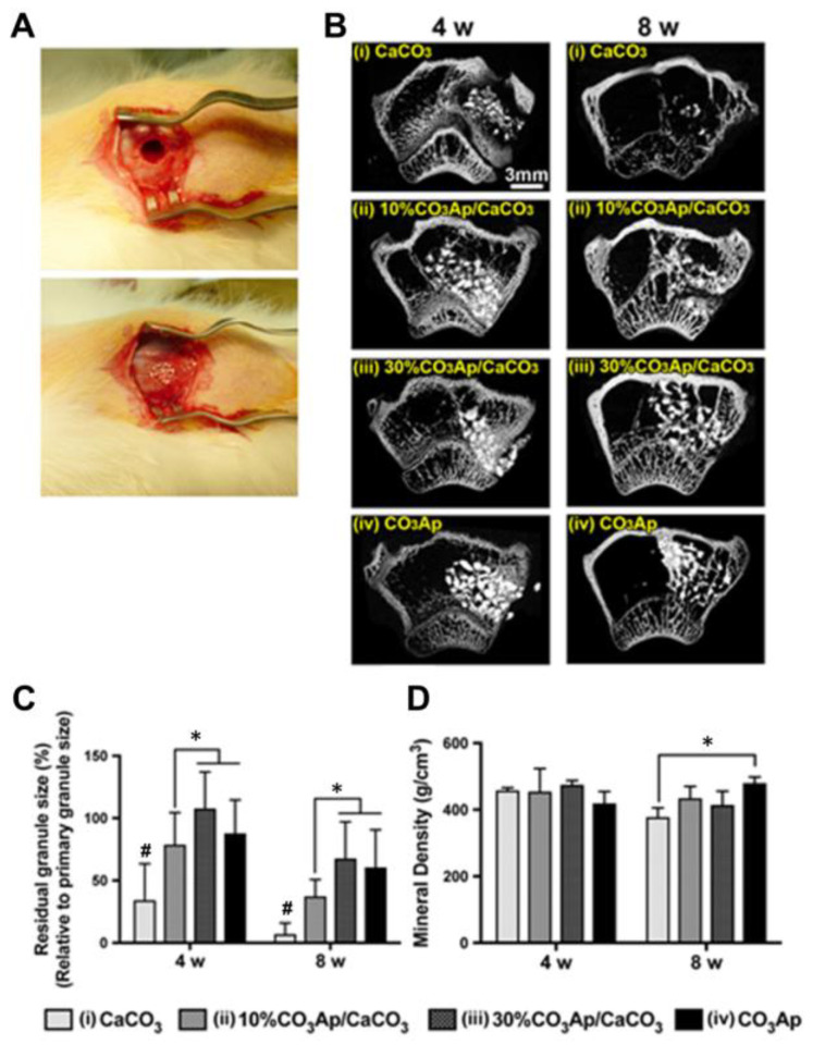
Keywords: CaCO3 particles; US imaging; anticancer therapy; antimicrobial therapy; ayer-by-layer assembly; biodegradation; buffering; calcite; calcium carbonate; calcium ions; carbon dioxide bubbles; cavitation; controlled release; degradation; dissolution; metastability; ossification; pH-sensitivity; recrystallization; resorption; theranostics; vaterite.


Conflict of interest statement

The authors declare no conflict of interest.

Figures

Figure 1
Figure 1
Incorporation of the drug molecules within the vaterite particles by adsorption and co-precipitation methods at room temperature (RT), and via freezing induced loading at −20 °C.
Figure 2
Figure 2
(A) Schematic representation of the vaterite–calcite recrystallization process. Reproduced with permission from [101]. (B) Schematics for the process of drug liberation from vaterite carriers, which is mediated by the vaterite–calcite recrystallization. Reproduced from Open Access Article [107]. (C) Release of water-insoluble drugs from vaterite carriers in aqueous media resulting in the formation of insoluble crystals (particles) by payload molecules. SEM images and results of EDX analysis illustrating the degradation process of the carriers loaded with griseofulvin (Gf) antifungal drug in deionized water. The precipitated Gf particles are contoured with orange. Reproduced with permission from [43]. (D) Release of water-soluble drugs from vaterite carriers in aqueous media. Schematics of the release process and corresponding two-photon fluorescence microscopy and SEM (insets) images of the carriers loaded with rhodamine 6G before and after their incubation in water. Adapted with permission from [33].
Figure 3
Figure 3
Intracellular recrystallization of vaterite carriers. Schematics of the vaterite–calcite transformation (the upper row), CLSM images of HeLa cells after their incubation with the carriers for 10 min, 3 and 24 h (the middle row) and the results of Raman analysis of a single cell after 72 h incubation with the carriers (the bottom row). Adapted with permission from [111].
Figure 4
Figure 4
Exploiting of Ca2+ release, which occurs during the vaterite–calcite recrystallization, for improvement of the ossification process in vitro. (A) Schematic representation of the cellular treatment using vaterite carriers loaded with alkaline phosphatase (ALP). (B) Live cells stained by calcein AM (green), (C) fixed cells stained by the Osteoimage mineralization assay (Cyan) and (D) hydroxyapatite deposition measured at different times using the Osteoimage mineralization assay on MC3T3-E1 cells. Reproduced with permission from [123].
Figure 5
Figure 5
Recrystallization-driven drug release from the vaterite carriers in vivo. Schematic representation of tannic acid (TA) release from the vaterite-coated polycaprolactone (PCL) fibers subcutaneously implanted in rats (the upper row). Schematics and SEM images illustrating the process of the vaterite-coating formation and its transformation to calcite in an aqueous medium in vitro (the middle row). SEM images and results of EDX analysis illustrating the vaterite–calcite transformation of the fiber coating in vivo (the lower row). Reproduced with permission from [7].
Figure 6
Figure 6
pH-dependent dissolution of vaterite carriers triggering the payload release. (A) Schematic representation of the decomposition-mediated drug release process from the vaterite carriers depending on the pH of the medium. Reproduced with permission from [38]. (B) SEM images of micro- and submicron vaterite carriers loaded with a photosensitizer before their incubation in various media and the phase-schemes illustrating the process of their transformation in acetate buffers (pH 4.5–6.5) and in water (pH 7.0) during 24 h. Reproduced with permission from [38]. (C) Schematics and SEM images illustrating the dissolution of vaterite-based carriers (VHC) and kinetics of the pH-dependent release of the loaded doxorubicin (DOX) drug. Reproduced with permission from [97].
Figure 7
Figure 7
Exploiting pH-sensitivity of vaterite carriers for drug delivery to tumors. (A) Schematic presentation of vaterite application in tumor targeting. Reproduced from Open Access Article [18]. (B) An example of successful application of the vaterite particles loaded with a porphyrazine (pz) drug and gold nanorods (GNR) in photodynamic therapy of tumors. Adapted with permission from [98].
Figure 8
Figure 8
Prevention of the burst release from vaterite carriers via their surface modification. (A) Effect of the layer-by-layer (LbL) coating formation on the BSA release from the vaterite carriers: schematics of the polyelectrolyte layers deposition, CLSM image of the LbL-coated carriers and BSA release kinetics from the bare and coated carriers at different pH (6.5 and 7.4). Adapted with permission from [44,127,140]. (B) Effect of carboxymethyl cellulose (CMC) incorporation and further coating of the vaterite matrices with chitosan/alginate (Chi/Alg) multilayers on the payload release: schematics, SEM-image and kinetics of DOX liberation from the CMC-doped carriers, both coated and non-coated with Chi/Alg, at pH 5.0. Adapted with permission from [78].
Figure 9
Figure 9
Enhancement of the vaterite carriers targeting to tumors via their surface modification. (A) Schematics representing the structure of vaterite carriers loaded with Mn2+-chelated chlorin e6 photosensitizer (Ce6(Mn)) and modified with PEG. (B) Schematic illustration of the pH-responsive decomposition of the carriers (incubation in PBS at pH 5.5, 6.5 and 7.4). (C) pH-triggered MR enhancement and MR-imaging monitored photosensitizer release in vitro. Adapted with permission from [141].
Figure 10
Figure 10
Schematic illustration for fabrication of the DOX-loaded calcium carbonate-crosslinked polypeptide carriers (CaNP/DOX), their circulation in vivo, intratumoral accumulation and pH-triggered intracellular DOX release. Reproduced from Open Access Article [142].
Figure 11
Figure 11
Vaterite–nanosilver hybrids with antibacterial properties and pH-triggered release. (A) SEM images and surface roughness plots of the pristine (CaCO3/AgNPs) and PSS-modified (CaCO3-PSS/AgNPs) vaterite–nanosilver hybrids. (B) Cumulative release of silver ions from the hybrids in PBS at pH 5.0, 7.4 and 9.0. (C) Schematic illustration of the AgNPs release driven by the recrystallization and dissolution of the hybrids. Adapted from Open Access Article [147].
Figure 12
Figure 12
Exploiting of Ca2+ release, which occurs during the dissolution of vaterite particles, for triggering the alginate gelation (A) and accelerating the blood clotting (B). (A) reproduced with permission from [55], (B) from [149].
Figure 13
Figure 13
Exploiting of CO2 bubbles’ generation, which occurs during the dissolution of vaterite particles, for US imaging of tumors. Schematic illustration of the pH-dependent CO2 generation and the images showing the US contrast in tumor, liver and subcutaneous area after the injection of DOX-loaded vaterite carriers in vivo to the corresponding site. Reproduced with permission from [152].
Figure 14
Figure 14
Schematic of the pH/ultrasound dual-responsive CO2 generation for US imaging-guided therapeutic inertial cavitation and sonodynamic therapy. (A) Formation of the hematoporphyrin monomethyl ether (HMME)-loaded vaterite carriers coated with hyaluronic acid (HA). (B,C) Mechanism of the tumor destruction utilizing the carriers and US treatment. Reproduced with permission from [153].
Figure 15
Figure 15
In vivo degradation of the CaCO3-based implanting material for improvement of ossification in bone tissue engineering. (A) Implantation of the fabricated granules in cylindrical bone defects of the rabbit femur. (B) Horizontal μCT views of the rabbit femur defect with CaCO3, 10% CO3Ap/CaCO3, 30% CO3Ap/CaCO3 and CO3Ap granules at 4 and 8 weeks. (C) The residual granules area (%) quantified by μCT at 4 and 8 weeks. (D) Mineral density the bone defect area at 4 and 8 weeks. An asterisk (*) denotes significant differences between groups, p < 0.05; a hash (#) denotes significantly lower than all other modalities, p < 0.05. Reproduced with permission from [161].
Figure 16
Figure 16
The scheme of the micro- and nanoparticle internalization by endocytic mechanism and intracellular transport. Reproduced with permission from [169].
Figure 17
Figure 17
In vivo degradation of vaterite carriers in hair follicles. (A) SEM, CLSM images and schematics illustrating the intrafollicular delivery of the carriers. Reproduced with permission from [170]. (B) SEM (the left column) and CLSM (the middle and right columns) images illustrating the process of the carriers’ degradation inside the hair follicles of rats in vivo. (C) Excretion kinetics of the fluorescent dye intrafollicularly delivered by means of degradable vaterite carries. (B) and (C) reproduced with permission from [16].
Figure 18
Figure 18
Prolongation of in vivo degradation of the vaterite carriers and sustainment of the payload release via formation of the stabilizing coating on the carriers’ surface. (A) Schematics illustrating the formation of vaterite carriers loaded with a griseofulvin (Gf) drug and coated with poly-L-arginine (PA), dextran sulfate (DS) and heparin (HP) polyelectrolytes. (B) Schematics of the Gf urinary excretion rate investigation. (C) Urinary excretion profiles of Gf after its administration by means of (Gf-CaCO3) and (Gf-CaCO3)/(PA/DS)2/HP carriers or after pure Gf application in rats in vivo. An asterisk (*) indicates significant differences in the Gf peak intensity at a particular time point after drug delivery as compared to the control urine value (zero time point) within the same group (p < 0.05). An ampersand (&) shows a significant difference in the Gf peak intensities between the group of (Gf-CaCO3)/(PA/DS)2/HP carriers and the pure Gf group (p < 0.05) on the last day of the experiment. Reproduced with permission from [43].
Figure 19
Figure 19
Stabilization of metastable vaterite in CaCO3 biomineralization through the addition of ovalbumin protein. (AC) Calcium carbonate particles formed in the presence of 0.2 gL−1 ovalbumin. (D) XRD spectrum of the CaCO3-ovalbumin precipitates, where “C” indicates calcite peaks (JCPDS: 05-0586), “V”—vaterite ones (JCPDS: 33-0268). Scale bars correspond to 1 μm (A,B) and 100 nm (C,D). Reproduced with permission from [26].
Figure 20
Figure 20
CLSM and SEM images of carboxymethyl–dextran–FITC/vaterite hybrids (A) and diethylaminoethyl–dextran–FITC/vaterite hybrids (B). Scale bar is 10 µm for (i), (ii) and (iii), 1 µm for (iv), and 100 nm for (v).Reproduced with permission from [175].
Figure 21
Figure 21
Scheme illustrating the process of vaterite stabilization by mucin incorporation. Reproduced with permission from [83].
Figure 22
Figure 22
Schematics of possible mechanisms of CaCO3 biomineralization. Reproduced with permission from [187].

Similar articles

Cited by

References

    1. Liendo F., Arduino M., Deorsola F.A., Bensaid S. Factors Controlling and Influencing Polymorphism, Morphology and Size of Calcium Carbonate Synthesized through the Carbonation Route: A Review. Powder Technol. 2022;398:117050. doi: 10.1016/j.powtec.2021.117050. - DOI
    1. Trushina D.B., Borodina T.N., Belyakov S., Antipina M.N. Calcium Carbonate Vaterite Particles for Drug Delivery: Advances and Challenges. Mater. Today Adv. 2022;14:100214. doi: 10.1016/j.mtadv.2022.100214. - DOI - PMC - PubMed
    1. Ferreira A.M., Vikulina A.S., Volodkin D. CaCO3 Crystals as Versatile Carriers for Controlled Delivery of Antimicrobials. J. Control. Release. 2020;328:470–489. doi: 10.1016/j.jconrel.2020.08.061. - DOI - PubMed
    1. Huang Y., Cao L., Parakhonskiy B.V., Skirtach A.G. Hard, Soft, and Hard-and-Soft Drug Delivery Carriers Based on CaCO3 and Alginate Biomaterials: Synthesis, Properties, Pharmaceutical Applications. Pharmaceutics. 2022;14:909. doi: 10.3390/pharmaceutics14050909. - DOI - PMC - PubMed
    1. Boyjoo Y., Pareek V.K., Liu J. Synthesis of Micro and Nano-Sized Calcium Carbonate Particles and Their Applications. J. Mater. Chem. A. 2014;2:14270–14288. doi: 10.1039/C4TA02070G. - DOI

LinkOut - more resources