. 2023 Oct 26;11(11):2632.
doi: 10.3390/microorganisms11112632.

In Silico Evaluation, Phylogenetic Analysis, and Structural Modeling of the Class II Hydrophobin Family from Different Fungal Phytopathogens

Affiliations

In Silico Evaluation, Phylogenetic Analysis, and Structural Modeling of the Class II Hydrophobin Family from Different Fungal Phytopathogens

Nahla A Bouqellah et al. Microorganisms. .

Abstract

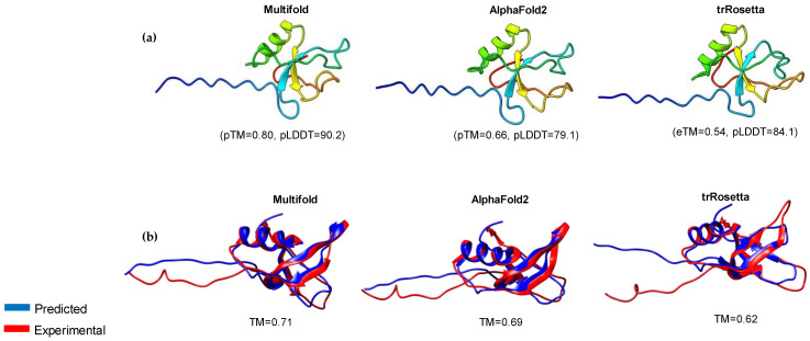
The class II hydrophobin group (HFBII) is an extracellular group of proteins that contain the HFBII domain and eight conserved cysteine residues. These proteins are exclusively secreted by fungi and have multiple functions with a probable role as effectors. In the present study, a total of 45 amino acid sequences of hydrophobin class II proteins from different phytopathogenic fungi were retrieved from the NCBI database. We used the integration of well-designed bioinformatic tools to characterize and predict their physicochemical parameters, novel motifs, 3D structures, multiple sequence alignment (MSA), evolution, and functions as effector proteins through molecular docking. The results revealed new features for these protein members. The ProtParam tool detected the hydrophobicity properties of all proteins except for one hydrophilic protein (KAI3335996.1). Out of 45 proteins, six of them were detected as GPI-anchored proteins by the PredGPI server. Different 3D structure templates with high pTM scores were designed by Multifold v1, AlphaFold2, and trRosetta. Most of the studied proteins were anticipated as apoplastic effectors and matched with the ghyd5 gene of Fusarium graminearum as virulence factors. A protein-protein interaction (PPI) analysis unraveled the molecular function of this group as GTP-binding proteins, while a molecular docking analysis detected a chitin-binding effector role. From the MSA analysis, it was observed that the HFBII sequences shared conserved 2 Pro (P) and 2 Gly (G) amino acids besides the known eight conserved cysteine residues. The evolutionary analysis and phylogenetic tree provided evidence of episodic diversifying selection at the branch level using the aBSREL tool. A detailed in silico analysis of this family and the present findings will provide a better understanding of the HFBII characters and evolutionary relationships, which could be very useful in future studies.

Keywords: computational annotation; effectors; evolution; homology modeling; hydrophobins.


Conflict of interest statement

The authors declare no conflict of interest.

Figures

Figure 1
Figure 1
Physicochemical characteristics of the hydrophobin proteins: (a) protein length vs. GRAVY scores, where the negative values were categorized as globular (hydrophilic) proteins while the positive values were categorized as membrane (hydrophobic) proteins; (b) theoretical isoelectric point (PI) of hydrophobin proteins; and (c) hydropathy plot of Ustulina deusta cerato-ulmin HFBII.
Figure 2
Figure 2
The number of GPI-anchored and non-GPI-anchored HFBII proteins with illustrated schematic diagram about GPI-anchoring localization outside the membrane.
Figure 3
Figure 3
Homology modeling of representative HFBII protein: (a) three-dimensional models of Verticillium dahlia protein (XP_009650899.1) were generated using MultiFold, AlphaFold2, and trRosetta, showing TM-scores and pLDDT values; (b) structural superposition between the experimental (PDB: 4AOG) and predicted structures for the selected HFBII protein.
Figure 4
Figure 4
Three-dimensional (3D) models of other representative fungal proteins that resemble HFBII proteins, with different pTM scores in Multifold v1.
Figure 5
Figure 5
Model validation of protein (XP_009650899.1) and two-dimensional structure prediction: (a) B-factor coloring, indicating the protein residue quality; (b) protein model evaluation using ModFOLD8, representing the confidence and p-value; (c) schematic and topology diagram showing the secondary structural elements in the protein; and (d) comparative method, including five tools for predicting the 2D structure of HFBII proteins using the Quick2D server and visualization with 2dSS.
Figure 6
Figure 6
(a) Bar graph illustrating the effector and non-effector HFBII proteins; (b) STRING PPI network analysis between representative query HFBII (XP_009650899.1) and GTP-binding proteins. The average node degree is 5.6, the average local clustering coefficient is 0.778, and the PPI enrichment p-value is 5.28 × 10⁻⁵.
Figure 7
Figure 7
Domain and intrinsic disorder protein analysis: (a) domain profile of 8 selected HFBII proteins, illustrating a mutant bacterial domain in the KAI3335996.1 protein; (b) the prediction of the disordered regions for the hydrophobin II fusion protein with a pentapeptide domain; (c) conservation patterns for the KAI3335996.1 protein across several phytopathogen HFBII proteins that show a highly variable, disordered middle region (pentapeptide domain); and (d) the prediction of the disordered regions for the hydrophobin II representative protein without the pentapeptide fusion part.
Figure 8
Figure 8
The conserved profile from alignment sequences of the selected HFBII proteins showed the twelve conserved residues (8 Cys, 2 Pro, and 2 Gly). The yellow color at the conservation bar below the figure indicates the 100% conservation residues.
Figure 9
Figure 9
Construction of phylogenetic tree by MEGA 11 and visualization via iTol v6. Motif locations were identified using the MEME server.
Figure 10
Figure 10
Positive selection analysis of the HFBII proteins using the Selecton server.
Figure 11
Figure 11
FUBAR and aBSREL evolutionary analyses: (a) FUBAR analysis of a coding sequence alignment to determine whether some sites have been subject to pervasive purifying or diversifying selection; (b) omega (ω) distribution over node 38 from the phylogenetic analysis using the aBSREL web server; and (c) omega (ω) distribution over a Microdochium trichocladiopsis node from the phylogenetic analysis using the aBSREL web server.
Figure 12
Figure 12
Active site information of an HFBII protein (XP_009650899.1): (a) eight cavities, detected by the scfbio server in the active site; (b) the amino acid residues (blue color) in the active site of the studied protein that were detected by the CASTp server.
Figure 13
Figure 13
Molecular docking modeling between chitin oligomer (ligand) and (a) beta-N-acetylglucosaminidase (receptor); (b) experimental hydrophobin (receptor); and (c) predicted hydrophobin (receptor).

References

    1. Li X., Wang F., Xu Y., Liu G., Dong C. Cysteine-rich hydrophobin gene family: Genome wide analysis, phylogeny and transcript Profiling in Cordyceps militaris. Int. J. Mol. Sci. 2021;22:643. doi: 10.3390/ijms22020643. - DOI - PMC - PubMed
    1. Bayry J., Aimanianda V., Guijarro J.I., Sunde M., Latge´ J.P. Hydrophobins—Unique fungal proteins. PLoS Pathog. 2012;8:e1002700. doi: 10.1371/journal.ppat.1002700. - DOI - PMC - PubMed
    1. Quarantin A., Hadeler B., Kröger C., Schäfer W., Favaron F., Sella L., Martínez-Rocha A.L. Different hydrophobins of Fusarium graminearum are involved in hyphal growth, attachment, water-Air interface penetration and plant infection. Front. Microbiol. 2019;10:751. doi: 10.3389/fmicb.2019.00751. - DOI - PMC - PubMed
    1. Vergunst K.L., Kenward C., Langelaan D.N. Characterization of the structure and self-assembly of two distinct class IB hydrophobins. Appl. Microbiol. Biotechnol. 2022;106:7831–7843. doi: 10.1007/s00253-022-12253-x. - DOI - PubMed
    1. Tanaka T., Terauchi Y., Yoshimi A., Abe K. Aspergillus hydrophobins: Physicochemical properties, biochemical properties, and functions in solid polymer degradation. Microorganisms. 2022;10:1498. doi: 10.3390/microorganisms10081498. - DOI - PMC - PubMed

LinkOut - more resources