

Extracellular Vesicles Secreted by Acanthamoeba culbertsoni Have COX and Proteolytic Activity and Induce Hemolysis
- PMID: 38004773
- PMCID: PMC10673465
- DOI: 10.3390/microorganisms11112762
Extracellular Vesicles Secreted by Acanthamoeba culbertsoni Have COX and Proteolytic Activity and Induce Hemolysis
Abstract
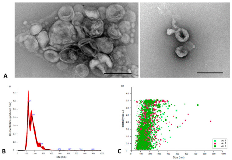
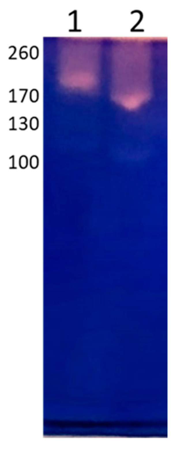
Several species of Acanthamoeba genus are potential pathogens and etiological agents of several diseases. The pathogenic mechanisms carried out by these amoebae in different target tissues have been documented, evidencing the relevant role of contact-dependent mechanisms. With the purpose of describing the pathogenic processes carried out by these protozoans more precisely, we considered it important to determine the emission of extracellular vesicles (EVs) as part of the contact-independent pathogenicity mechanisms of A. culbertsoni, a highly pathogenic strain. Through transmission electronic microscopy (TEM) and nanoparticle tracking analysis (NTA), EVs were characterized. EVs showed lipid membrane and a size between 60 and 855 nm. The secretion of large vesicles was corroborated by confocal and TEM microscopy. The SDS-PAGE of EVs showed proteins of 45 to 200 kDa. Antigenic recognition was determined by Western Blot, and the internalization of EVs by trophozoites was observed through Dil-labeled EVs. In addition, some EVs biological characteristics were determined, such as proteolytic, hemolytic and COX activity. Furthermore, we highlighted the presence of leishmanolysin in trophozites and EVs. These results suggest that EVs are part of a contact-independent mechanism, which, together with contact-dependent ones, allow for a better understanding of the pathogenicity carried out by Acanthamoeba culbertsoni.
Keywords: Acanthamoeba culbertsoni; COX; extracellular vesicles; pathogenicity mechanisms.
Conflict of interest statement
The authors declare no conflict of interest.
Figures


Similar articles
-
Advances in the study of extracellular vesicles of Naegleria fowleri and their role in contact-independent pathogenic mechanisms.Parasit Vectors. 2025 May 1;18(1):164. doi: 10.1186/s13071-025-06786-z. Parasit Vectors. 2025. PMID: 40312759 Free PMC article.
-
Comprehensive characterization of extracellular vesicles produced by environmental (Neff) and clinical (T4) strains of Acanthamoeba castellanii.mSystems. 2024 Jun 18;9(6):e0122623. doi: 10.1128/msystems.01226-23. Epub 2024 May 8. mSystems. 2024. PMID: 38717186 Free PMC article.
-
Cytopathic changes in rat microglial cells induced by pathogenic Acanthamoeba culbertsoni: morphology and cytokine release.Clin Diagn Lab Immunol. 2001 Jul;8(4):837-40. doi: 10.1128/CDLI.8.4.837-840.2001. Clin Diagn Lab Immunol. 2001. PMID: 11427438 Free PMC article.
-
The Methods of Choice for Extracellular Vesicles (EVs) Characterization.Int J Mol Sci. 2017 May 29;18(6):1153. doi: 10.3390/ijms18061153. Int J Mol Sci. 2017. PMID: 28555055 Free PMC article. Review.
-
Pathogenic free-living amoebae.Kisaengchunghak Chapchi. 1990 Dec;28 Suppl:29-39. doi: 10.3347/kjp.1990.28.suppl.29. Kisaengchunghak Chapchi. 1990. PMID: 2133420 Review.
Cited by
-
Advances in the study of extracellular vesicles of Naegleria fowleri and their role in contact-independent pathogenic mechanisms.Parasit Vectors. 2025 May 1;18(1):164. doi: 10.1186/s13071-025-06786-z. Parasit Vectors. 2025. PMID: 40312759 Free PMC article.
-
Detection of immunogenic protein components in excretion/secretion products of Acanthamoeba T5 using polyclonal antibodies.Mem Inst Oswaldo Cruz. 2025 Jun 9;120:e240190. doi: 10.1590/0074-02760240190. eCollection 2025. Mem Inst Oswaldo Cruz. 2025. PMID: 40498906 Free PMC article.
-
Extracellular vesicles isolated from SphK1 inhibitor SKI II-medium restrain the migration of colorectal cancer.Transl Cancer Res. 2025 May 30;14(5):2594-2602. doi: 10.21037/tcr-24-2152. Epub 2025 May 7. Transl Cancer Res. 2025. PMID: 40530117 Free PMC article.
-
Clusterin-mediated polarization of M2 macrophages: a mechanism of temozolomide resistance in glioblastoma stem cells.Stem Cell Res Ther. 2025 Mar 24;16(1):146. doi: 10.1186/s13287-025-04247-z. Stem Cell Res Ther. 2025. PMID: 40128761 Free PMC article.
-
Comprehensive characterization of extracellular vesicles produced by environmental (Neff) and clinical (T4) strains of Acanthamoeba castellanii.mSystems. 2024 Jun 18;9(6):e0122623. doi: 10.1128/msystems.01226-23. Epub 2024 May 8. mSystems. 2024. PMID: 38717186 Free PMC article.
References
-
- Reddy D.L., van den Berg E., Grayson W., Mphahlele M., Frean J. Clinical Improvement of Disseminated Acanthamoeba Infection in a Patient with Advanced HIV Using a Non-Miltefosine-Based Treatment Regimen in a Low-Resource Setting. Trop. Med. Infect. Dis. 2022;7:24. doi: 10.3390/tropicalmed7020024. - DOI - PMC - PubMed
Grants and funding
LinkOut - more resources
Full Text Sources
