. 2023 Nov 15;11(11):2777.
doi: 10.3390/microorganisms11112777.

Potent Antiviral Activity of Vitamin B12 against Severe Acute Respiratory Syndrome Coronavirus 2, Middle East Respiratory Syndrome Coronavirus, and Human Coronavirus 229E

Affiliations

Potent Antiviral Activity of Vitamin B12 against Severe Acute Respiratory Syndrome Coronavirus 2, Middle East Respiratory Syndrome Coronavirus, and Human Coronavirus 229E

Yassmin Moatasim et al. Microorganisms. .

Abstract

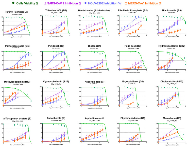
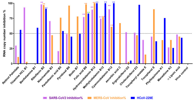
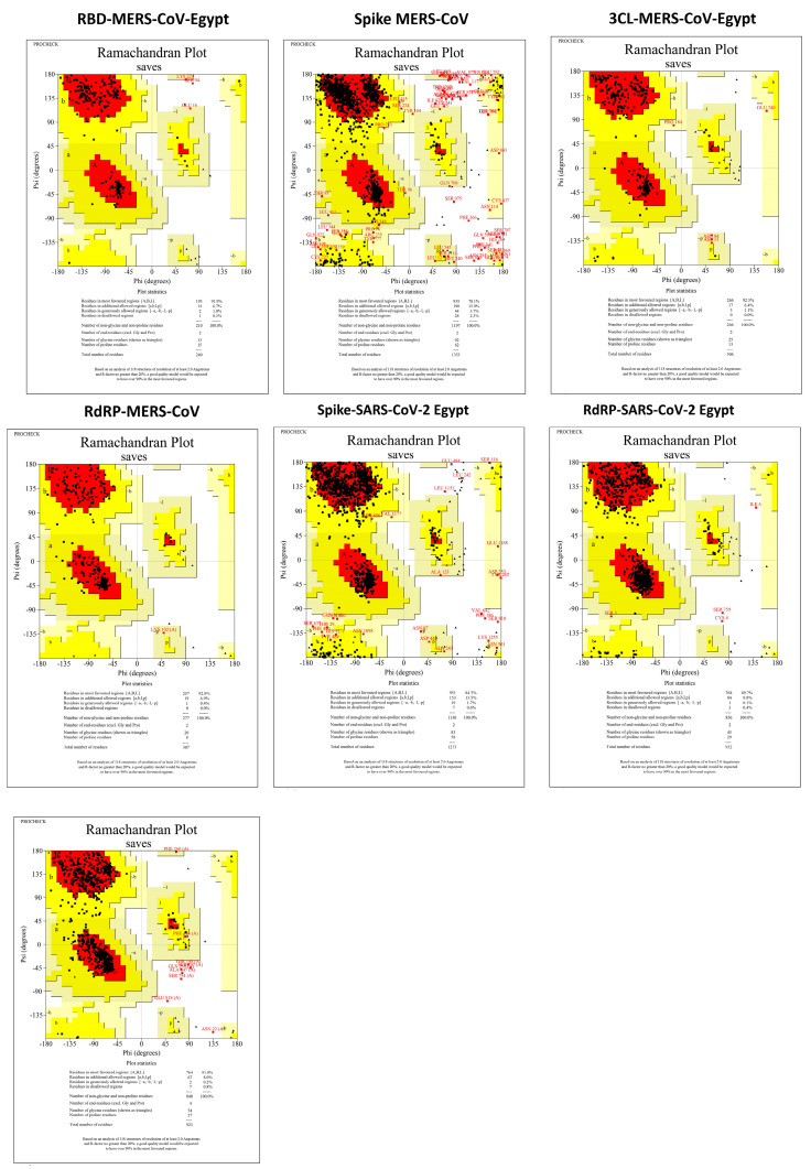
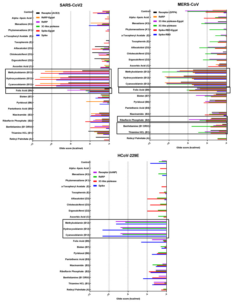
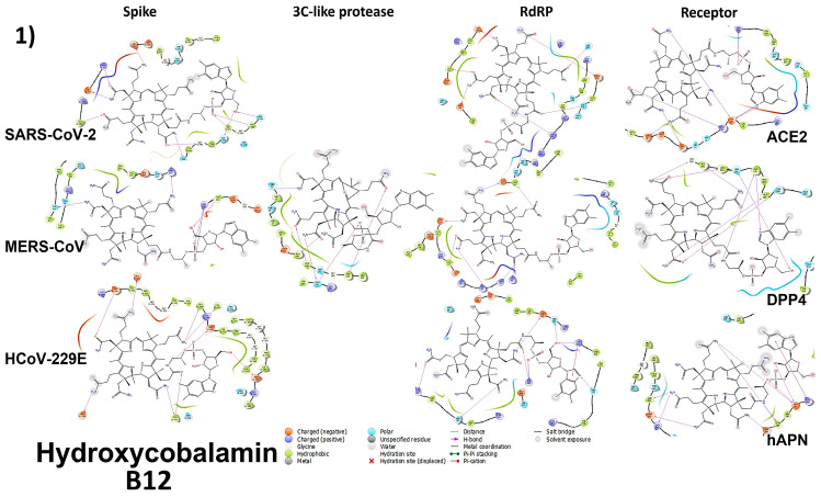
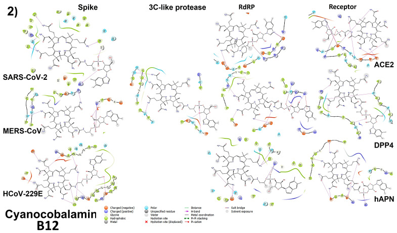
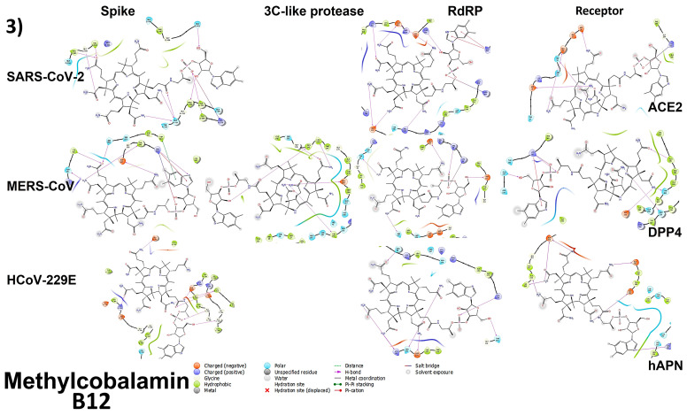
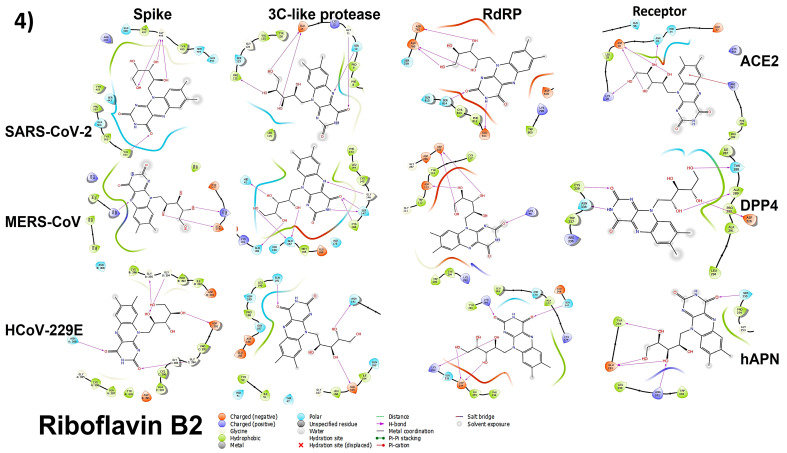
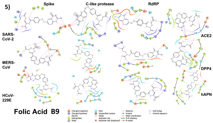
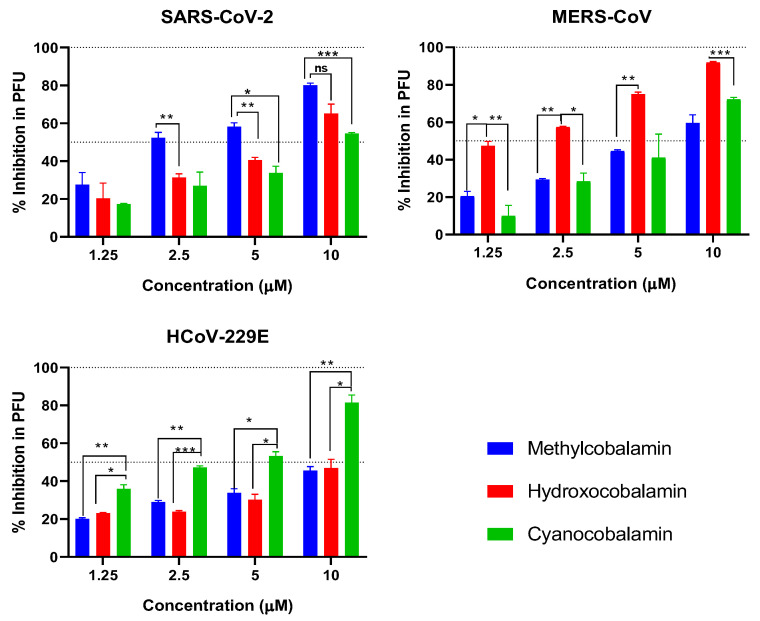
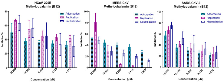
Repurposing vitamins as antiviral supporting agents is a rapid approach used to control emerging viral infections. Although there is considerable evidence supporting the use of vitamin supplementation in viral infections, including severe acute respiratory syndrome coronavirus 2 (SARS-CoV-2), the specific role of each vitamin in defending against coronaviruses remains unclear. Antiviral activities of available vitamins on the infectivity and replication of human coronaviruses, namely, SARS-CoV-2, Middle East respiratory syndrome coronavirus (MERS-CoV), and human coronavirus 229E (HCoV-229E), were investigated using in silico and in vitro studies. We identified potential broad-spectrum inhibitor effects of Hydroxocobalamin and Methylcobalamin against the three tested CoVs. Cyanocobalamin could selectively affect SARS-CoV-2 but not MERS-CoV and HCoV-229E. Methylcobalamin showed significantly higher inhibition values on SARS-CoV-2 compared with Hydroxocobalamin and Cyanocobalamin, while Hydroxocobalamin showed the highest potent antiviral activity against MERS-CoV and Cyanocobalamin against HCoV-229E. Furthermore, in silico studies were performed for these promising vitamins to investigate their interaction with SARS-CoV-2, MERS-CoV, and HCoV-229E viral-specific cell receptors (ACE2, DPP4, and hAPN protein, respectively) and viral proteins (S-RBD, 3CL pro, RdRp), suggesting that Hydroxocobalamin, Methylcobalamin, and Cyanocobalamin may have significant binding affinity to these proteins. These results show that Methylcobalamin may have potential benefits for coronavirus-infected patients.

Keywords: MERS-CoV; SARS-CoV-2; antiviral agent; viral infection; vitamin B12; vitamins.


Conflict of interest statement

The authors declare no conflict of interest.

Figures

Figure 1
Figure 1
Two-dimensional chemical structures of vitamins included in this study as obtained from PubChem web server.
Figure 2
Figure 2
Determination of CC50 and IC50 of vitamins in Vero E6 cells against SARS-CoV-2, MERS-CoV, and HCoV-229E. Values of CC50 and IC50 were calculated using non-linear regression analysis with Graph Pad Prism software (version 5.01) by plotting log inhibitor versus normalized response (variable slope); All values are listed in Table 1.
Figure 3
Figure 3
Viral inhibition percentage of viral RNA copy numbers of SARS-CoV-2, MERS-CoV, and HCoV-229E at 24 h post-infection and treatment with vitamins using Vero E6 cells (MOI 0.05). Real-time RT-PCR was used to compare the treatment with untreated virus control. Small stars represent the level of significance compared with the virus control tested using GraphPad prism software v7 (2-way ANOVA test with Dunnett’s post-test and 95% confidence level). Colors of stars match the color of specific column. Significance values are represented as follows, ** refers to p ≤ 0.01.
Figure 4
Figure 4
Ramachandran plot showing the phi/psi values of the modeled proteins of the three coronaviruses. Based on analyses of 118 structures with resolution of at least 2.0 angstroms and R—factor no greater than 20%, a good quality model was expected to have over 90% in the most favored regions. Red colorfavorable regions are represented by red color, while the yellow color is the allowed region, and white color is the disallowed regions. Black dots are the amino acid residues.
Figure 5
Figure 5
Binding energies (as a Glide score) of the tested vitamins against different proteins correlated with the three coronaviruses. All score values are in negative charge. The space between the bars in each vitamin means no binding was detected or binding energies were higher than −4 kcal/mol (less stable binding). Black boxes indicate the lowest, i.e., strongest, binding. Egypt in protein names indicates the 3D modeling protein from sequences of isolated MERS and SARS-2 viruses from Egypt involved in this study in in vitro experiments, while other proteins were retrieved as 3D structures from the PDB as reference strains.
Figure 6
Figure 6
Molecular docking of the 3 forms of B12 vitamin (13), B2 (4), and B9 (5) on the viral proteins and cell receptors of SARS-CoV-2, MERS-CoV, and HCoV-229E. Horizontal axes represent the viruses and the vertical axes represent the proteins. Empty spots represent no binding. Purple lines represent the formation of Hydrogen bonds. Red and blue residues are charged ones, light blue are polar ones, and green ones are hydrophobic.
Figure 6
Figure 6
Molecular docking of the 3 forms of B12 vitamin (13), B2 (4), and B9 (5) on the viral proteins and cell receptors of SARS-CoV-2, MERS-CoV, and HCoV-229E. Horizontal axes represent the viruses and the vertical axes represent the proteins. Empty spots represent no binding. Purple lines represent the formation of Hydrogen bonds. Red and blue residues are charged ones, light blue are polar ones, and green ones are hydrophobic.
Figure 7
Figure 7
Plaque reduction assay of three tested forms of vitamin B12, namely, Methylcobalamin (blue), hydroxocobalamin (red), and cyanocobalamin (green), against SARS-CoV-2, MERS-CoV, and 229E viruses at doses of 1.25-2.5-5-10 micromoles. Statistical analyses were performed using two-way ANOVA, followed by Bonferroni’s multiple comparisons test, where the confidence interval was set to 95%. * refers to p ≤ 0.05, ** refers to p ≤ 0.01, *** refers to p ≤ 0.001.
Figure 8
Figure 8
Mode of action of Methylcobalamin against the three coronaviruses on VERO-E6 cells, showing that vitamin b12 reduced viral replication in different stages in viral replication cycle of the three tested coronaviruses.

Similar articles

Cited by

References

    1. Dong E., Du H., Gardner L. An interactive web-based dashboard to track COVID-19 in real time. Lancet Infect. Dis. 2020;20:533–534. doi: 10.1016/S1473-3099(20)30120-1. - DOI - PMC - PubMed
    1. Graham R.L., Donaldson E.F., Baric R.S. A decade after SARS: Strategies for controlling emerging coronaviruses. Nat. Rev. Microbiol. 2013;11:836–848. doi: 10.1038/nrmicro3143. - DOI - PMC - PubMed
    1. Su S., Wong G., Shi W., Liu J., Lai A.C.K., Zhou J., Liu W., Bi Y., Gao G.F. Epidemiology, Genetic Recombination, and Pathogenesis of Coronaviruses. Trends Microbiol. 2016;24:490–502. doi: 10.1016/j.tim.2016.03.003. - DOI - PMC - PubMed
    1. Gaunt E.R., Hardie A., Claas E.C., Simmonds P., Templeton K.E. Epidemiology and Clinical Presentations of the Four Human Coronaviruses 229E, HKU1, NL63, and OC43 Detected over 3 Years Using a Novel Multiplex Real-Time PCR Method. J. Clin. Microbiol. 2010;48:2940–2947. doi: 10.1128/JCM.00636-10. - DOI - PMC - PubMed
    1. Lim Y.X., Ng Y.L., Tam J.P., Liu D.X. Human Coronaviruses: A Review of Virus–Host Interactions. Diseases. 2016;4:26. doi: 10.3390/diseases4030026. - DOI - PMC - PubMed