. 2023 Nov 17;11(11):2801.
doi: 10.3390/microorganisms11112801.

Regulatory Functions of PurR in Yersinia pestis: Orchestrating Diverse Biological Activities

Affiliations

Regulatory Functions of PurR in Yersinia pestis: Orchestrating Diverse Biological Activities

Liting Xiao et al. Microorganisms. .

Abstract

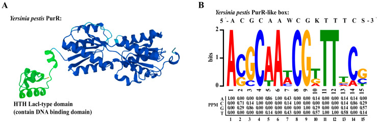
The bacterium Yersinia pestis has developed various strategies to sense and respond to the complex stresses encountered during its transmission and pathogenic processes. PurR is a common transcriptional regulator of purine biosynthesis among microorganisms, and it modulates the transcription level of the pur operon to suppress the production of hypoxanthine nucleotide (IMP). This study aims to understand the functions and regulatory mechanisms of purR in Y. pestis. Firstly, we constructed a purR knockout mutant of Y. pestis strain 201 and compared certain phenotypes of the null mutant (201-ΔpurR) and the wild-type strain (201-WT). The results show that deleting purR has no significant impact on the biofilm formation, growth rate, or viability of Y. pestis under different stress conditions (heat and cold shock, high salinity, and hyperosmotic pressure). Although the cytotoxicity of the purR knockout mutant on HeLa and 293 cells is reduced, the animal-challenging test found no difference of the virulence in mice between 201-ΔpurR and 201-WT. Furthermore, RNA-seq and EMSA analyses demonstrate that PurR binds to the promoter regions of at least 15 genes in Y. pestis strain 201, primarily involved in purine biosynthesis, along with others not previously observed in other bacteria. Additionally, RNA-seq results suggest the presence of 11 potential operons, including a newly identified co-transcriptional T6SS cluster. Thus, aside from its role as a regulator of purine biosynthesis, purR in Y. pestis may have additional regulatory functions.

Keywords: Yersinia pestis; purR; purine biosynthesis; transcriptional regulation.


Conflict of interest statement

The authors declare no conflict of interest.

Figures

Figure 1
Figure 1
The growth curves of 201-WT and 201-ΔpurR. The growth curves of 201-WT and 201-ΔpurR were assessed under different culture conditions. The conditions included: (A) growth at 26 °C in LB medium, (B) growth at 37 °C in LB medium, (C) growth at 26 °C in TMH medium, and (D) growth at 37 °C in TMH medium. The bar graph presented below the growth curves illustrates the cumulative areas under the curves and is applied to statistical analysis. Each experiment included three independent biological replicates, and the results were expressed as mean ± standard deviation from three independent experiments. ns: not statistically significant.
Figure 2
Figure 2
The in vitro phenotypes of 201-WT and 201-ΔpurR. Biofilm formation of 201-WT and 201-ΔpurR and a comparison of their survival rates in vitro under different simulated stress environments were assessed. (A) 0.1% crystal violet solution was used to quantify the relative amount of biofilm formation for both strains cultured at 26 °C or 37 °C (B). The survival rates of both strains after being stimulated by high osmotic pressure environment simulated by 0.5 M sorbitol after 30 min and after 1.5 h were compared. The survival rates of both strains after being stimulated by heat shock at 50 °C for 0.5 h (C) and cold shock at 4 °C for 24 h (D) were compared. (E) The survival rates of both strains after being stimulated by high salt environment simulated by 7.5% NaCl were compared after 1 h of stimulation. There were no significant differences in all results between 201-WT and 201-ΔpurR. Each experiment was independently replicated three times for both strains, and statistical analysis was performed using a two-sample t-test for each comparison.
Figure 3
Figure 3
201. purR showed cytotoxicity attenuation on HeLa cells and 293 cells. Cells were infected with 201-WT, 201-ΔpurR, and 201-ΔpurR-Comp at a specific multiplicity of infection (MOI). (A) Bacterial infection of HeLa cells at an MOI of 5. (B) Bacterial infection of 293 cells at an MOI of 5. (C) Bacterial infection of HeLa cells at an MOI of 10. (D) Bacterial infection of 293 cells at an MOI of 10. The cell index was measured every 15 min. The bar graph presented next to the curves illustrates the cumulative areas under the curves and is applied to statistical analysis. At the same time point, a larger “Normalized cell index” indicates more cells or a better cell status. The higher the curve, the larger the area under the curve, indicating that the bacterial strain has weaker cytotoxicity to the cells. Each experiment included three independent biological replicates, and the results were expressed as mean ± standard deviation from three independent experiments. * p < 0.05, ** p < 0.01, **** p < 0.0001, ns: not statistically significant.
Figure 4
Figure 4
Common differentially expressed genes of 201-ΔpurR cultured at 26 °C and 37 °C. The transcriptional level of 201-ΔpurR was analyzed using RNA-seq and qRT-PCR under 26 °C and 37 °C culture conditions. (A) The 21 up-regulated or down-regulated genes that were shared under 26 °C and 37 °C culture conditions, screened with the criterion of |log2(FoldChange)| > 1.0; all the selected genes had a p-adjust value of < 10−5. (B) The correlation analysis between the 17 genes selected for qRT-PCR and these same 17 genes in the RNA-seq under 26 °C culture conditions, and the figure took point (2,2) as the origin. The selected genes were listed in the Section 2.9.
Figure 5
Figure 5
PurR structure and prediction of PurR motif. (A) The structure of Y. pestis PurR on Uniprot. Green color is the HTH lacl-type domain which contains DNA-binding domain in the front of PurR. (B) PurR motif predicted on the online MEME website, based on the promoter region of six genes (purH, purE, purT, purL, purF, purM) on the pur operon.
Figure 6
Figure 6
PurR may regulate some operons involved in purine biosynthesis of Y. pestis strain 201. The results of EMSA and RT-PCR experiments confirm the presence of operons and the regulation by PurR in Y. pestis strain 201, involving genes related to purine biosynthesis. (A) The figure illustrates the experimental setup and schematic representation of each lane in the EMSA experiment. The filled black color represents the result of the experimental group, and the shaded grid indicates the result of the negative control group. The concentrations of the components added in each experimental channel are shown in Supplementary Table S2. The width of the bands reflects the quantity of binding between the tracer probe and PurR, while the number of ‘+’ signs corresponds to the amount of the respective samples added. The identified operons include: (B) purM-purN; (C) purH-purD; (D) purE-purK; and (E) guaB-guaA. The left figure displays the EMSA results, while the right figure presents the RT-PCR results. In the RT-PCR results, the template for each gene intergenic region is indicated as DNA, RNA, cDNA, or water. The blue dotted line indicates the expected amplification fragment size for each gene intergenic region.
Figure 7
Figure 7
Other operon of Y. pestis strain 201. The RT-PCR results revealed the co-transcription of specific genes in the genome of Y. pestis strain 201, which was supported with RNA-seq analysis (excluding previously mentioned genes). The co-transcribed regions identified include: (A) YP_RS00935-tauA-tauB-tauC-tauD and (B) the type VI secretion system (T6SS).

Similar articles

Cited by

References

    1. Yang R., Atkinson S., Chen Z., Cui Y., Du Z., Han Y., Sebbane F., Slavin P., Song Y., Yan Y., et al. Yersinia pestis and Plague: Some knowns and unknowns. Zoonoses. 2023;3:5. doi: 10.15212/ZOONOSES-2022-0040. - DOI - PMC - PubMed
    1. Valles X., Stenseth N.C., Demeure C., Horby P., Mead P.S., Cabanillas O., Ratsitorahina M., Rajerison M., Andrianaivoarimanana V., Ramasindrazana B., et al. Human plague: An old scourge that needs new answers. PLoS Neglected Trop. Dis. 2020;14:e0008251. doi: 10.1371/journal.pntd.0008251. - DOI - PMC - PubMed
    1. Li B., Tan Y., Guo J., Cui B., Wang Z., Wang H., Zhou L., Guo Z., Zhu Z., Du Z., et al. Use of protein microarray to identify gene expression changes of Yersinia pestis at different temperatures. Can. J. Microbiol. 2011;57:287–294. doi: 10.1139/w11-007. - DOI - PubMed
    1. Achtman M., Zurth K., Morelli G., Torrea G., Guiyoule A., Carniel E. Yersinia pestis, the cause of plague, is a recently emerged clone of Yersinia pseudotuberculosis. Proc. Natl. Acad. Sci. USA. 1999;96:14043–14048. doi: 10.1073/pnas.96.24.14043. - DOI - PMC - PubMed
    1. Achtman M. Evolution, population structure, and phylogeography of genetically monomorphic bacterial pathogens. Annu. Rev. Microbiol. 2008;62:53–70. doi: 10.1146/annurev.micro.62.081307.162832. - DOI - PubMed

LinkOut - more resources