. 2023 Nov 20;11(11):2818.
doi: 10.3390/microorganisms11112818.

Notch Signaling Regulates the Function and Phenotype of Dendritic Cells in Helicobacter pylori Infection

Affiliations

Notch Signaling Regulates the Function and Phenotype of Dendritic Cells in Helicobacter pylori Infection

Qiaoyuan Liu et al. Microorganisms. .

Abstract

Notch signaling manipulates the function and phenotype of dendritic cells (DCs), as well as the interaction between DCs and CD4+ T cells. However, the role of Notch signaling in Helicobacter pylori (H. pylori) infection remains elusive. Murine bone marrow-derived dendritic cells (BMDCs) were pretreated in the absence or presence of Notch signaling inhibitor DAPT prior to H. pylori stimulation and the levels of Notch components, cytokines and surface markers as well as the differentiation of CD4+ T cells in co-culture were measured using quantitative real-time PCR (qRT-PCR), Western blot, enzyme-linked immunosorbent assay (ELISA) and flow cytometry. Compared with the control, the mRNA expression of all Notch receptors and Notch ligands Dll4 and Jagged1 was up-regulated in H. pylori-stimulated BMDCs. The blockade of Notch signaling by DAPT influenced the production of IL-1β and IL-10 in H. pylori-pulsed BMDCs, and reduced the expression of Notch1, Notch3, Notch4, Dll1, Dll3 and Jagged2. In addition, DAPT pretreatment decreased the expression of maturation markers CD80, CD83, CD86, and major histocompatibility complex class II (MHC-II) of BMDCs, and further skewed Th17/Treg balance toward Treg. Notch signaling regulates the function and phenotype of DCs, thus mediating the differentiation of CD4+ T cells during H. pylori infection.

Keywords: CD4+ T cells; Helicobacter pylori; Notch signaling; dendritic cells.


Conflict of interest statement

The authors declare no conflict of interest.

Figures

Figure 1
Figure 1
Expression profile of Notch molecules in BMDCs during H. pylori infection. BMDCs were stimulated with H. pylori (MOI 50) for 24 h. PBS was used as control for H. pylori. qRT-PCR was performed to assess the mRNA expression of (A) Notch receptors and (B) Notch ligands in BMDCs. Relative expression is normalized to β-Actin. (C) Western blot was performed to assess the protein levels of Dll1, Dll4 and Jagged1 in BMDCs. β-Tubulin was used as loading control. (D) BMDCs were stimulated with different MOI (5, 10, 20 and 50) of H. pylori for 8 h, 16 h or 24 h, and qRT-PCR was performed to assess the mRNA level of Jagged1. Flow cytometry was performed to measure the level of Jagged1 on BMDCs stimulated with H. pylori (MOI 20) for 16 h, and (E) the representative histogram plot and (F) the percentage were illustrated. The data are presented as the mean ± SD of three independent experiments. * p < 0.05, ** p < 0.01, *** p < 0.001, **** p < 0.0001, N.S: no statistical difference.
Figure 2
Figure 2
Inhibition of Notch signaling influenced the production of cytokines in BMDCs during H. pylori infection. BMDCs were pretreated with DAPT (20 μM) for 24 h, and then stimulated with H. pylori (MOI 50) for 24 h. DMSO was used as control for DAPT. (A) ELISA was used to determine the level of IL-1β, IL-6, TNF-α and IL-10. qRT-PCR was performed to examine the mRNA expression of (B) Notch receptors and (C) Notch ligands. Relative expression is normalized to β-Actin. The data are presented as the mean ± SD of three independent experiments. * p < 0.05, ** p < 0.01 and *** p < 0.001, **** p < 0.0001, N.S: no statistical difference.
Figure 3
Figure 3
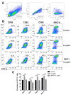
Inhibition of Notch signaling decreased the expression of maturation markers on BMDCs during H. pylori infection. BMDCs were pretreated with DAPT (20 μM) for 24 h, and then stimulated with H. pylori (MOI 50) for another 24 h. Naïve BMDCs were used as uninfected controls. DMSO was used as a control for DAPT. (A,B) Representative dot plots and the gating strategy of CD11c+ BMDCs are shown. Flow cytometry was performed to evaluate the expression of CD80, CD83, CD86 and MHC-II on BMDCs. (C) Percentages of BMDCs expressing CD80, CD83, CD86 or MHC-II are shown. The data are presented as the mean ± SD of three experiments. ** p < 0.01 and **** p < 0.0001.
Figure 4
Figure 4
The mRNA expression profile of Th cells was reversed by DAPT in the co-culture system of H. pylori-infected BMDCs and CD4+ T cells. BMDCs pretreated in the presence or absence of DAPT (20 μM, 24 h) were stimulated with H. pylori (MOI 50, 24 h) and then co-cultured with splenic CD4+ T cells from syngeneic C57BL/6 mice for 72 h. The mRNA expression of characteristic transcription factors and cytokines of (A) Th1 (Tbx21, Ifnγ), (B) Th2 (Gata3, Il4), (D) Th17 (Rorγt, Il17A, Il17F) and (E) Treg (Foxp3, Tgfβ, Il10) was assessed using qRT-PCR. (C) The ratio of Tbx21/Gata3 and (F) Rorγt/Foxp3 is shown. Relative expression is normalized to β-Actin. The data are presented as the mean ± SD of three independent experiments. * p < 0.05, ** p < 0.01 and *** p < 0.001, **** p < 0.0001, N.S: no statistical difference.
Figure 5
Figure 5
DAPT-pretreated BMDCs skewed Th17/Treg balance toward Treg. BMDCs pretreated in the presence or absence of DAPT (20 μM, 24 h) were stimulated with H. pylori (MOI 50, 24 h) and then co-cultured with splenic CD4+ T from syngeneic C57BL/6 mice for 72 h. Flow cytometric analyses were performed to measure the differentiation of CD4+ T cells. (A) Representative dot plots of Th1 (IFN-γ), Th2 (IL-4), Th17 (IL-17A) and Treg (Foxp3) are shown. (B) Percentages of Th1, Th2, Th17 and Treg and (C) the ratio of Th1/Th2 and Th17/Treg are shown. The data are presented as the mean ± SD of three independent experiments. * p < 0.05, ** p < 0.01, and N.S: no statistical difference.

Similar articles

Cited by

References

    1. Zamani M., Ebrahimtabar F., Zamani V., Miller W.H., Alizadeh-Navaei R., Shokri-Shirvani J., Derakhshan M.H. Systematic review with meta-analysis: The worldwide prevalence of Helicobacter pylori infection. Aliment. Pharmacol. Ther. 2018;47:868–876. doi: 10.1111/apt.14561. - DOI - PubMed
    1. Hooi J.K.Y., Lai W.Y., Ng W.K., Suen M.M.Y., Underwood F.E., Tanyingoh D., Malfertheiner P., Graham D.Y., Wong V.W.S., Wu J.C.Y., et al. Global Prevalence of Helicobacter pylori Infection: Systematic Review and Meta-Analysis. Gastroenterology. 2017;153:420–429. doi: 10.1053/j.gastro.2017.04.022. - DOI - PubMed
    1. Kao J.Y., Zhang M., Miller M.J., Mills J.C., Wang B., Liu M., Eaton K.A., Zou W., Berndt B.E., Cole T.S., et al. Helicobacter pylori Immune Escape Is Mediated by Dendritic Cell–Induced Treg Skewing and Th17 Suppression in Mice. Gastroenterology. 2010;138:1046–1054. doi: 10.1053/j.gastro.2009.11.043. - DOI - PMC - PubMed
    1. Constantino J., Gomes C., Falcão A., Neves B.M., Cruz M.T. Dendritic cell-based immunotherapy: A basic review and recent advances. Immunol. Res. 2017;65:798–810. doi: 10.1007/s12026-017-8931-1. - DOI - PubMed
    1. Shiu J., Blanchard T.G. Dendritic cell function in the host response to Helicobacter pylori infection of the gastric mucosa. Pathog. Dis. 2013;67:46–53. doi: 10.1111/2049-632X.12014. - DOI - PubMed