Review
. 2023 Nov 7;28(22):7456.
doi: 10.3390/molecules28227456.

Advancing Our Understanding of Pyranopterin-Dithiolene Contributions to Moco Enzyme Catalysis

Affiliations
Review

Advancing Our Understanding of Pyranopterin-Dithiolene Contributions to Moco Enzyme Catalysis

Sharon J Nieter Burgmayer et al. Molecules. .

Abstract

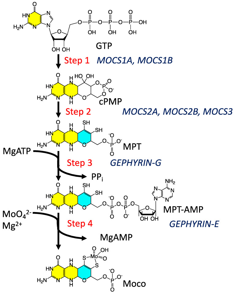
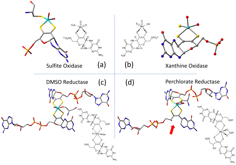
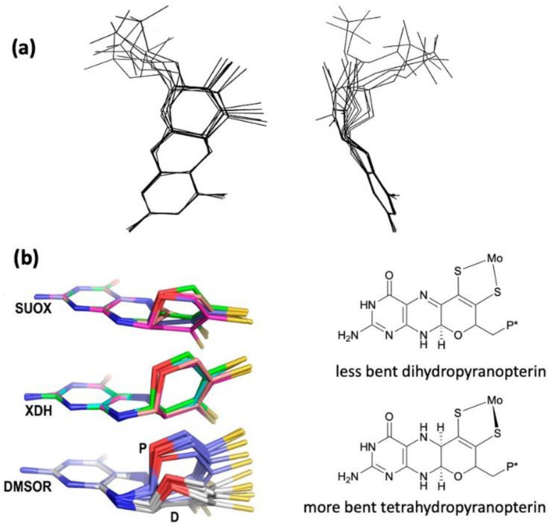
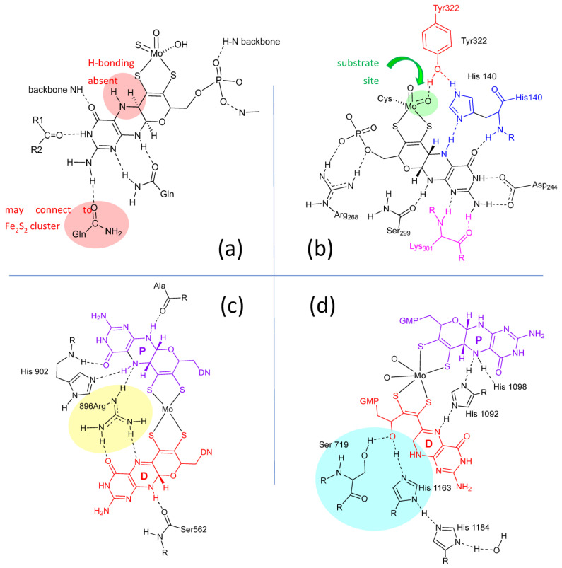
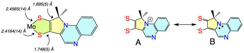
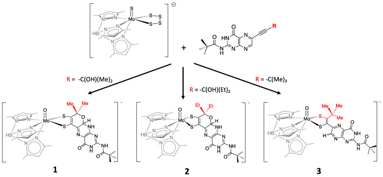
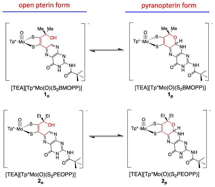
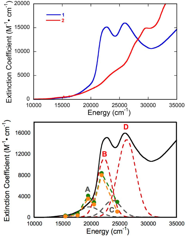
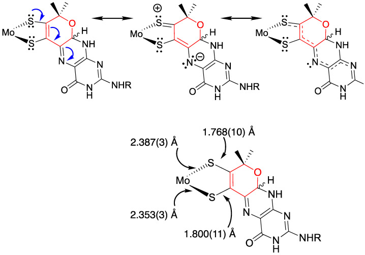
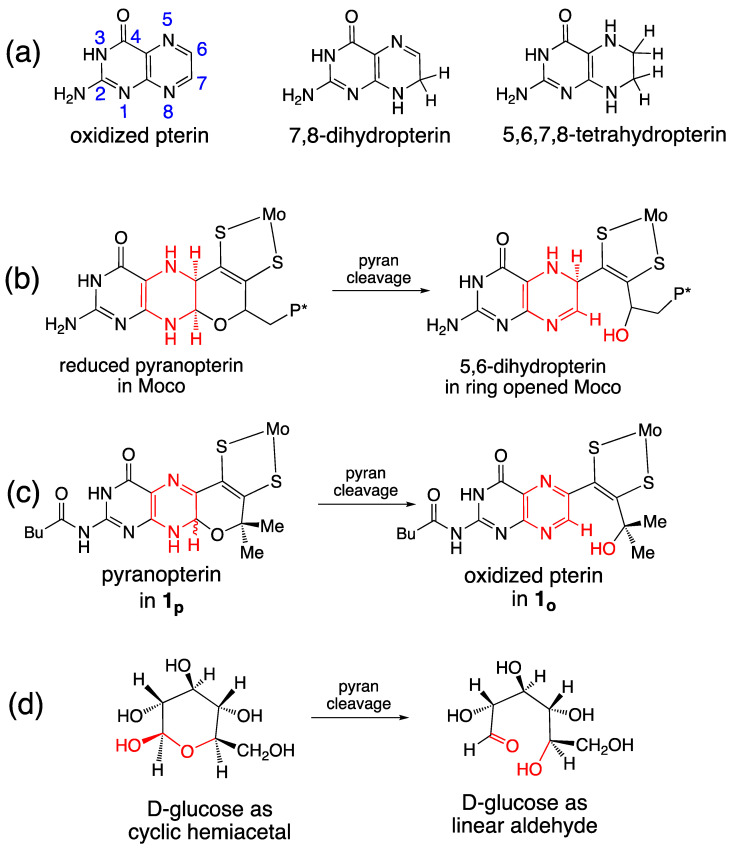
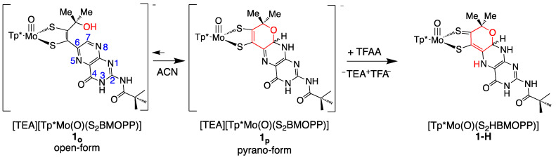
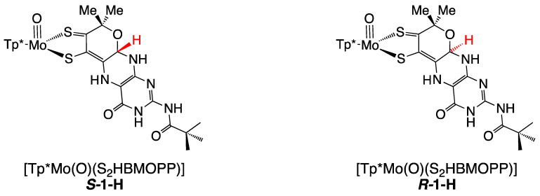
The pyranopterin dithiolene ligand is remarkable in terms of its geometric and electronic structure and is uniquely found in mononuclear molybdenum and tungsten enzymes. The pyranopterin dithiolene is found coordinated to the metal ion, deeply buried within the protein, and non-covalently attached to the protein via an extensive hydrogen bonding network that is enzyme-specific. However, the function of pyranopterin dithiolene in enzymatic catalysis has been difficult to determine. This focused account aims to provide an overview of what has been learned from the study of pyranopterin dithiolene model complexes of molybdenum and how these results relate to the enzyme systems. This work begins with a summary of what is known about the pyranopterin dithiolene ligand in the enzymes. We then introduce the development of inorganic small molecule complexes that model aspects of a coordinated pyranopterin dithiolene and discuss the results of detailed physical studies of the models by electronic absorption, resonance Raman, X-ray absorption and NMR spectroscopies, cyclic voltammetry, X-ray crystallography, and chemical reactivity.

Keywords: Moco; dithiolene; molybdenum cofactor; molybdenum enzymes; molybdopterin; pyranopterin.


Conflict of interest statement

The authors declare no conflict of interest.

Figures

Figure 1
Figure 1
The Moco biosynthetic pathway. The two yellow-highlighted rings comprise the pterin structure, while the cyan ring is the adjoining pyran ring.
Figure 2
Figure 2
Representative examples of Moco structures for each of the SO, XO, and DMSO families, and one example of Moco possessing one PDT having an open, uncyclized pyran ligand. (a) Sulfite Oxidase (PDB 1SOX). (b) Xanthine Oxidase (PDB 3NRZ). (c) Dimethylsulfoxide Reductase (PDB 1EU1). (d) Perchlorate Reductase (PDB 5CH7) where the red arrow points to the open pyran ring position.
Figure 3
Figure 3
(a) Two views of the range of pyranopterin conformations observed in 1997. (b) Two distinct conformations suggest that pyranopterins in Moco have different oxidation states in different families. P* denotes a phosphate or a dinucleotide terminus. Adapted from Ref. [79].
Figure 4
Figure 4
H-bonding interactions between the pyranopterin of Moco and adjacent protein residues. (a) Conserved H-bonding interactions identified for all 7 members of the XDH family. (b) Conserved H-bonding interactions identified for three members of the SUOX family. (c) H-bonding interactions within the DMSOR protein structure that are representative of 15 other members of the DMSOR family. (d) H-bonding interaction in E. coli nitrate reductase, whose Moco exhibits one non-cyclized pterin structure.
Figure 5
Figure 5
Low-frequency resonance Raman spectra for wt R. capsulatus XDH, Q197A and Q102G variants. Adapted with permission from Ref. [90]. Dong, C.; Yang, J.; Reschke, S.; Leimkühler, S.; Kirk, M.L. Vibrational Probes of Molybdenum Cofactor–Protein Interactions in Xanthine Dehydrogenase. Inorg. Chem. 2017, 56, 6830–6837. https://doi.org/10.1021/acs.inorgchem.7b00028. Copyright 2017 American Chemical Society.
Figure 6
Figure 6
Results from experiments probing the redox state of the pterin in Moco. Enzymes oxidized by either ferrocyanide or DCIP cause inactivation where Moco possesses an oxidized pterin in PDT (top). Oxidation stoichiometry implies active Moco is at the dihydropterin level of reduction having several possible tautomeric structures (bottom).
Figure 7
Figure 7
Redox reactivity of a model pyranopterin.
Figure 8
Figure 8
Top: Tp*MoO(dithiolene) first-generation model complexes that have been extensively probed spectroscopically using a combination of MCD, electronic absorption, photoelectron, electron paramagnetic resonance, and resonance Raman spectroscopies. Bottom: Symmetry coordinates for two totally symmetric low-frequency normal modes and their respective linear combinations.
Figure 9
Figure 9
(left) Bond lengths determined from X-ray crystallography for Tp*MoO(pyrrolo-S2BMOQO). (right) Contributing resonance structures for the ligand showing dominant dithiolene (A) and thione-thiolate (B) structures.
Figure 10
Figure 10
Pterin-dithiolene model compounds for Moco.
Figure 11
Figure 11
Model compounds [Tp*MoO(S2BMOPP)]1− (1) and [Tp*MoO(S2PEOPP)]1− (2) spontaneously cyclize, forming a pyran ring yielding the pyranopterin dithiolene structure found in Moco, and demonstrate pyran ring-chain tautomerization. In contrast, model compound [Tp*MoO(S2BDMPP)]1− (3) cannot form a pyran ring.
Figure 12
Figure 12
Electronic absorption spectra (298 K in DMSO) of [Tp*MoO(S2BMOPP)]1− (1) and ring-opened [Tp*MoO(S2BDMPP)]1−. Bands A–D derive from a Gaussian resolution of the electronic absorption spectrum. Adapted with permission from Ref. [111]. Gisewhite, D.R.; Yang, J.; Williams, B.R.; Esmail, A.; Stein, B.; Kirk, M.L.; Burgmayer, S.J.N. Implications of Pyran Cyclization and Pterin Conformation on Oxidized Forms of the Molybdenum Cofactor. J. Am. Chem. Soc. 2018, 140, 12808–12818. https://doi.org/10.1021/jacs.8b05777. Copyright 2018 American Chemical Society.
Figure 13
Figure 13
Contributing resonance structures leading to π delocalization in Moco, as evidenced by the bond alternation patterns in the dithiolene chelate ring. Adapted with permission from Ref. [111]. Gisewhite, D.R.; Yang, J.; Williams, B.R.; Esmail, A.; Stein, B.; Kirk, M.L.; Burgmayer, S.J.N. Im-plications of Pyran Cyclization and Pterin Conformation on Oxidized Forms of the Molybdenum Cofactor. J. Am. Chem. Soc. 2018, 140, 12808–12818. https://doi.org/10.1021/jacs.8b05777. Copyright 2018 American Chemical Society.
Figure 14
Figure 14
Pterin oxidation states and pyran ring opening outcomes for Moco and model compound 1. (a) Three oxidation levels of pterins. (b) Pyran ring opening in Moco. (c) Pyran ring opening in 1. (d) Ring-chain tautomerism in D-glucose.
Figure 15
Figure 15
Reaction of model compound 1 with trifluoroacetic acid. The dithiolene chelate is drawn as a partially oxidized thione/thiolate group based on experimental and computational data on the electronic structure of 1-H. Reproduced with permission from Ref. [92]. Gates, C.; Varnum, H.; Getty, C.; Loui, N.; Chen, J.; Kirk, M.L.; Yang, J.; Nieter Burgmayer, S.J. Protonation and Non-Innocent Ligand Behavior in Pyranopterin Dithiolene Molybdenum Com-plexes. Inorg. Chem. 2022, 61, 13728–13742. https://doi.org/10.1021/acs.inorgchem.2c01234. Copyright 2022 American Chemical Society.
Figure 16
Figure 16
DFT-optimized structures for R- and S-diastereomers of 1-H. Torsion angles between calculated dithiolene plane (atoms S-C=C-S) and pterin plane are 4.85 degrees for S-H7 and 2.80 degrees for R-H7. Reproduced with permission from Ref. [92]. Gates, C.; Varnum, H.; Getty, C.; Loui, N.; Chen, J.; Kirk, M.L.; Yang, J.; Nieter Burgmayer, S.J. Protonation and Non-Innocent Ligand Behavior in Pyranopterin Dithiolene Molybdenum Com-plexes. Inorg. Chem. 2022, 61, 13728–13742. https://doi.org/10.1021/acs.inorgchem.2c01234. Copyright 2022 American Chemical Society.
Figure 16
Figure 16
DFT-optimized structures for R- and S-diastereomers of 1-H. Torsion angles between calculated dithiolene plane (atoms S-C=C-S) and pterin plane are 4.85 degrees for S-H7 and 2.80 degrees for R-H7. Reproduced with permission from Ref. [92]. Gates, C.; Varnum, H.; Getty, C.; Loui, N.; Chen, J.; Kirk, M.L.; Yang, J.; Nieter Burgmayer, S.J. Protonation and Non-Innocent Ligand Behavior in Pyranopterin Dithiolene Molybdenum Com-plexes. Inorg. Chem. 2022, 61, 13728–13742. https://doi.org/10.1021/acs.inorgchem.2c01234. Copyright 2022 American Chemical Society.
Figure 17
Figure 17
Room-temperature electronic absorption spectra of 1 (red) and 1-H (purple) in ACN. Adapted with permission from Ref. [92]. Gates, C.; Varnum, H.; Getty, C.; Loui, N.; Chen, J.; Kirk, M.L.; Yang, J.; Nieter Burgmayer, S.J. Protonation and Non-Innocent Ligand Behavior in Pyranopterin Dithiolene Molybdenum Com-plexes. Inorg. Chem. 2022, 61, 13728–13742. https://doi.org/10.1021/acs.inorgchem.2c01234. Copyright 2022 American Chemical Society.
Figure 18
Figure 18
Correlation between the percent of thione-thiol resonance structure contribution to a series of model complexes, (AD), 1, and 1-H. The straight line was derived from a best fit of the data for organic thiols, thiones, and NBO computations. Reproduced with permission from Ref. [92]. Gates, C.; Varnum, H.; Getty, C.; Loui, N.; Chen, J.; Kirk, M.L.; Yang, J.; Nieter Burgmayer, S.J. Protonation and Non-Innocent Ligand Behavior in Pyranopterin Dithiolene Molybdenum Com-plexes. Inorg. Chem. 2022, 61, 13728–13742. https://doi.org/10.1021/acs.inorgchem.2c01234. Copyright 2022 American Chemical Society.
Figure 19
Figure 19
Protonated pyranopterin in 1-H facilitates redox reactions (1) and (2) with DCIP, O2, and DMSO that do not occur in the absence of protonated pyranopterin.
Figure 20
Figure 20
Comparison of pyran ring formation in pterin dithiolene and quinoxaline dithiolene compounds.
Figure 21
Figure 21
Protonation of 4 initiates pyran ring cyclization, forming unstable 4-H, followed by -loss of -OH and intramolecular cyclization to a pyrroloquinoxaline compound 5.
Figure 22
Figure 22
Bis-dithiolene oxo-Mo complexes with pyranoquinoxaline at different levels of reduction (oxidized, semi-reduced, reduced) photocatalyze reduction of H+ and CO2, where the proportion of reduced carbon products increases for the more reduced dithiolenes.
Figure 23
Figure 23
The electron transfer chain in XO, indicating a vectorial pathway for electron egress involving the Mo ion, the PDT, two spinach ferredoxin type 2Fe2S clusters, and a flavin. Electrons exit the enzyme at FAD.

Similar articles

Cited by

References

    1. Ingersol L.J., Kirk M.L. Structure, Function, and Mechanism of Pyranopterin Molybdenum and Tungsten Enzymes. In: Constable E.C., Parkin G., Que L. Jr., editors. Comprehensive Coordination Chemistry III. Elsevier; Oxford, UK: 2021. pp. 790–811.
    1. Yang J., Enemark J.H., Kirk M.L. Metal-Dithiolene Bonding Contributions to Pyranopterin Molybdenum Enzyme Reactivity. Inorganics. 2020;8:19. doi: 10.3390/inorganics8030019. - DOI - PMC - PubMed
    1. Kirk M.L., Kc K. Molybdenum and Tungsten Cofactors and the Reactions They Catalyze. Transition Metals and Sulfur—A Strong Relationship for Life. In: Sosa Torres M., Kroneck P., editors. Metal Ions in Life Sciences. Volume 20. De Gruyter; Berlin, Germany: 2020. pp. 313–342. - PMC - PubMed
    1. Hille R., Schulzke C., Kirk M.L. Molybdenum and Tungsten Enzymes. The Royal Society of Chemistry; Cambridge, UK: 2017.
    1. Kirk M.L., Hille R. Spectroscopic Studies of Mononuclear Molybdenum Enzyme Centers. Molecules. 2022;27:4802. doi: 10.3390/molecules27154802. - DOI - PMC - PubMed

LinkOut - more resources