. 2023 Nov 8;28(22):7477.
doi: 10.3390/molecules28227477.

The Phenolic Profile and Anti-Inflammatory Effect of Ethanolic Extract of Polish Propolis on Activated Human Gingival Fibroblasts-1 Cell Line

Affiliations

The Phenolic Profile and Anti-Inflammatory Effect of Ethanolic Extract of Polish Propolis on Activated Human Gingival Fibroblasts-1 Cell Line

Anna Kurek-Górecka et al. Molecules. .

Abstract

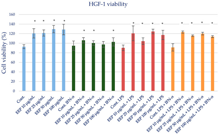
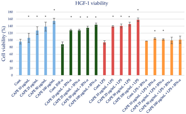
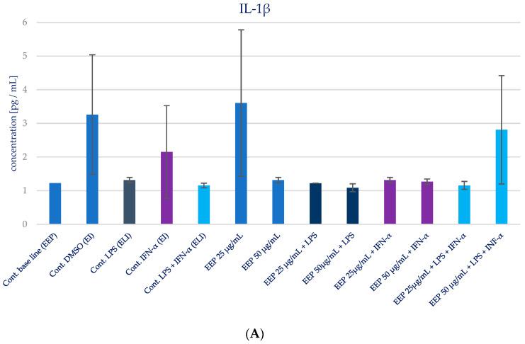
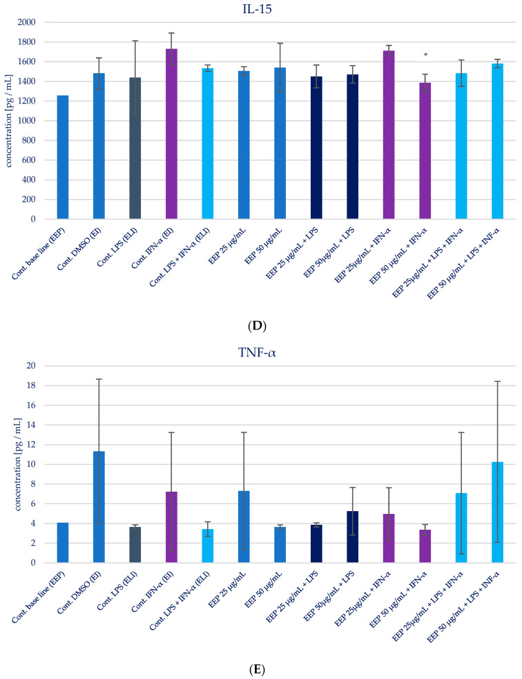
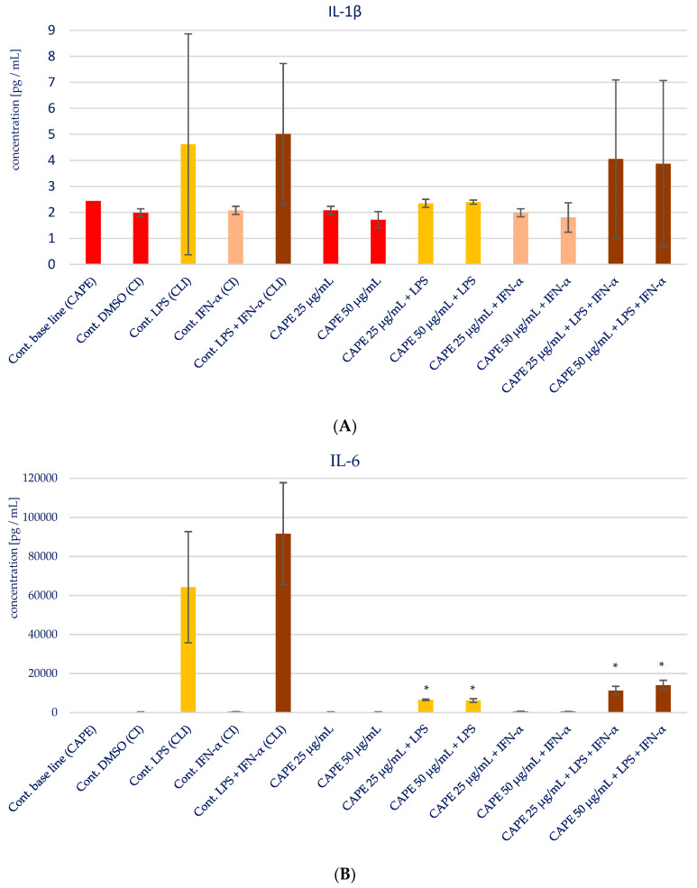
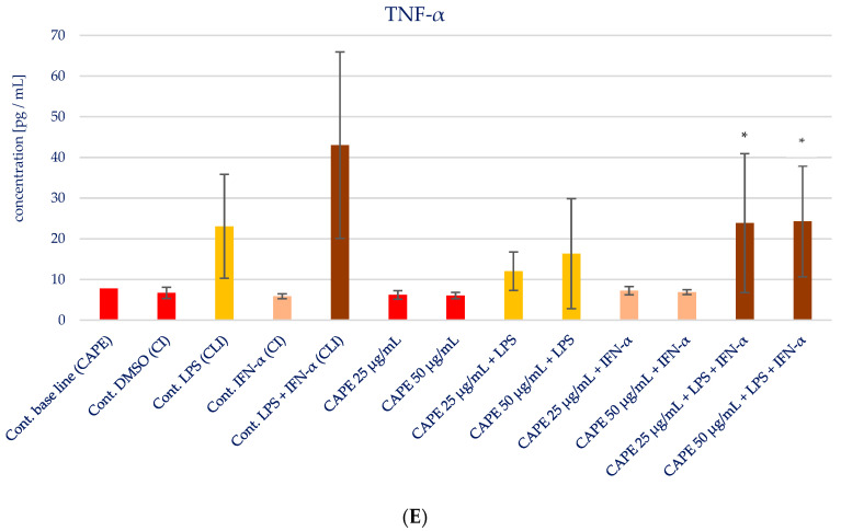
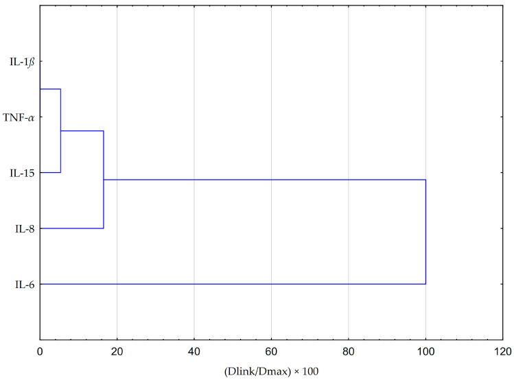
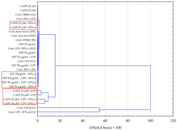
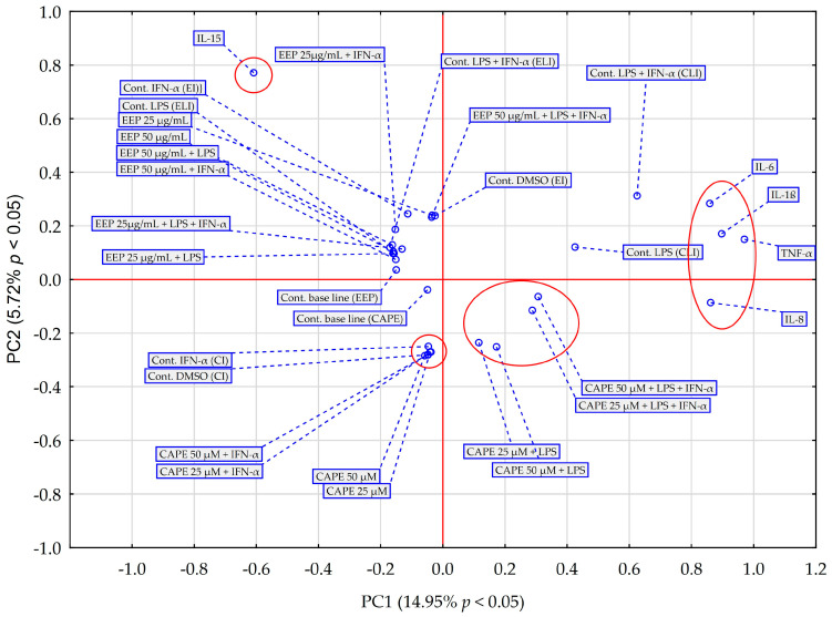
Propolis, owing to its antibacterial and anti-inflammatory properties, acts as a cariostatic agent, capable of preventing the accumulation of dental plaque and inhibiting inflammation. The anti-inflammatory properties of propolis are attributed to caffeic acid phenethyl ester (CAPE), which is present in European propolis. The objective of the conducted study was to assess the anti-inflammatory effects of the Polish ethanolic extract of propolis (EEP) and isolated CAPE on stimulated with LPS and IFN-α, as well as the combination of LPS and IFN-α. The cytotoxicity of the tested compounds was determined using the MTT assay. The concentrations of specific cytokines released by the HGF-1 cell line following treatment with EEP (25-50 µg/mL) or CAPE (25-50 µg/mL) were assessed in the culture supernatant. In the tested concentrations, both CAPE and EEP did not exert cytotoxic effects. Our results demonstrate that CAPE reduces TNF-α and IL-6 in contrast to EEP. Propolis seems effective in stimulating HGF-1 to release IL-6 and IL-8. A statistically significant difference was observed for IL-8 in HGF-1 stimulated by LPS+IFN-α and treated EEP at a concentration of 50 µg/mL (p = 0.021201). Moreover, we observed that CAPE demonstrates a stronger interaction with IL-8 compared to EEP, especially when CAPE was administered at a concentration of 50 µg/mL after LPS + IFN-α stimulation (p = 0.0005). Analysis of the phenolic profile performed by high-performance liquid chromatography allowed identification and quantification in the EEP sample of six phenolic acids, five flavonoids, and one aromatic ester-CAPE. Propolis and its compound-CAPE-exhibit immunomodulatory properties that influence the inflammatory process. Further studies may contribute to explaining the immunomodulatory action of EEP and CAPE and bring comprehensive conclusions.

Keywords: caffeic acid phenethyl ester; cytokines; in vitro model; inflammation; polyphenols; propolis.


Conflict of interest statement

The authors declare no conflict of interest.

Figures

Figure 1
Figure 1
Cell viability measured by MTT (%)—the cytotoxic activity of propolis ethanolic extract. The values represent the mean ± SD of three independent assays; * means p < 0.05 (calculated using t-test).
Figure 2
Figure 2
HGF-1 cells with CAPE incubated with MTT for 4 h presenting formazan crystals. (A). CAPE 10 µg/mL; (B). CAPE 25 µg/mL; (C). CAPE 50 µg/mL; (D). CAPE 100 µg/mL. (The arrow marks selected formazan crystals).
Figure 3
Figure 3
Cell viability measured by MTT (%)—the cytotoxic activity of CAPE. The values represent the mean ± SD of three independent assays; * means p < 0.05 (calculated using t-test).
Figure 4
Figure 4
Effect of EEP on selected cytokines production in native and stimulated HGF-1 by LPS, IFN-α, as well as combined LPS and IFN-α. (A). IL-1β, (B). IL-6, (C). IL-8, (D). IL-15, (E). TNF-α. The presented values mean ± SD of three independent experiments (n = 8); * means p < 0.05 (calculated using Fisher’s LSD Test).
Figure 4
Figure 4
Effect of EEP on selected cytokines production in native and stimulated HGF-1 by LPS, IFN-α, as well as combined LPS and IFN-α. (A). IL-1β, (B). IL-6, (C). IL-8, (D). IL-15, (E). TNF-α. The presented values mean ± SD of three independent experiments (n = 8); * means p < 0.05 (calculated using Fisher’s LSD Test).
Figure 5
Figure 5
Effect of CAPE on selected cytokines production in native and stimulated HGF-1 by LPS and IFN-α, as well as combined LPS and IFN-α; * means p < 0.05 (calculated using Fisher’s LSD Test).
Figure 5
Figure 5
Effect of CAPE on selected cytokines production in native and stimulated HGF-1 by LPS and IFN-α, as well as combined LPS and IFN-α; * means p < 0.05 (calculated using Fisher’s LSD Test).
Figure 5
Figure 5
Effect of CAPE on selected cytokines production in native and stimulated HGF-1 by LPS and IFN-α, as well as combined LPS and IFN-α; * means p < 0.05 (calculated using Fisher’s LSD Test).
Figure 6
Figure 6
Dendrogram obtained via the HCA analysis of all obtained data based on the average content of the effect of EEP and CAPE at different concentrations on pro-inflammatory cytokines.
Figure 7
Figure 7
Dendrogram obtained via the HCA analysis of all obtained data based on the influence on pro-inflammatory cytokines by the average content of the EEP and the CAPE at different concentrations.
Figure 8
Figure 8
PCA score plot of all obtained data based on the average content of the effect of EEP and CAPE at different concentrations on pro-inflammatory cytokines. PC—principal component.

Similar articles

Cited by

References

    1. Kurek-Górecka A., Walczyńska-Dragon K., Felitti R., Nitecka-Buchta A., Baron S., Olczyk P. The Influence of Propolis on Dental Plaque Reduction and the Correlation between Dental Plaque and Severity of COVID-19 Complications—A Literature Review. Molecules. 2021;26:5516. doi: 10.3390/molecules26185516. - DOI - PMC - PubMed
    1. Liccardo D., Cannavo A., Spagnuolo G., Ferrara N., Cittadini A., Rengo C., Rengo G. Periodontal Disease: A Risk Factor for Diabetes and Cardiovascular Disease. Int. J. Mol. Sci. 2019;20:1414. doi: 10.3390/ijms20061414. - DOI - PMC - PubMed
    1. Ceccarelli F., Saccucci M., Di Carlo G., Lucchetti R., Pilloni A., Pranno N., Luzzi V., Valesini G., Polimeni A. Periodontitis and Rheumatoid Arthritis: The Same Inflammatory Mediators? Mediat. Inflamm. 2019;2019:6034546. doi: 10.1155/2019/6034546. - DOI - PMC - PubMed
    1. Sansores-España D., Carrillo-Avila A., Melgar-Rodriguez S., Díaz-Zuñiga J., Martínez-Aguilar V. Periodontitis and alzheimer’s disease. Med. Oral. Patol. Oral. Cir. Bucal. 2021;26:e43–e48. doi: 10.4317/medoral.23940. - DOI - PMC - PubMed
    1. Gündogar H., Üstün K., Şenyurt S.Z., Özdemir E.Ç., Sezer U., Erciyas K. Gingival crevicular fluid levels of cytokine, chemokine, and growth factors in patients with periodontitis or gingivitis and periodontally healthy subjects: A cross-sectional multiplex study. Central Eur. J. Immunol. 2021;46:474–480. doi: 10.5114/ceji.2021.110289. - DOI - PMC - PubMed

LinkOut - more resources