. 2023 Nov 8;28(22):7484.
doi: 10.3390/molecules28227484.

Zaluzanin C Alleviates Inflammation and Lipid Accumulation in Kupffer Cells and Hepatocytes by Regulating Mitochondrial ROS

Affiliations

Zaluzanin C Alleviates Inflammation and Lipid Accumulation in Kupffer Cells and Hepatocytes by Regulating Mitochondrial ROS

Ji-Won Jung et al. Molecules. .

Abstract

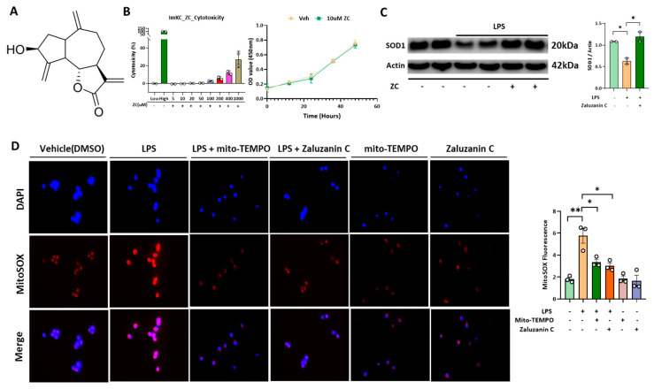
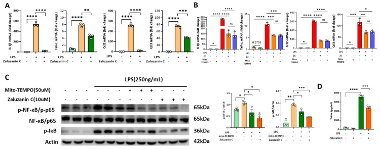
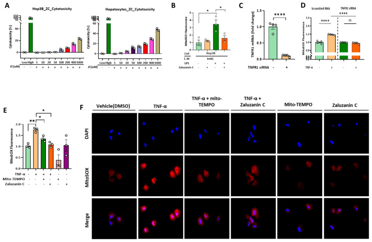
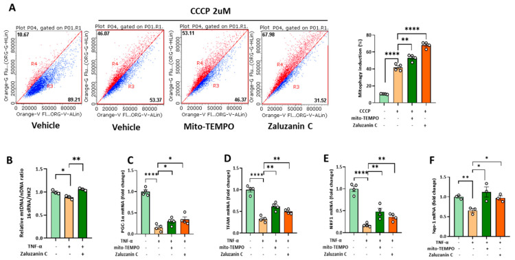
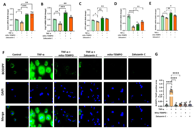
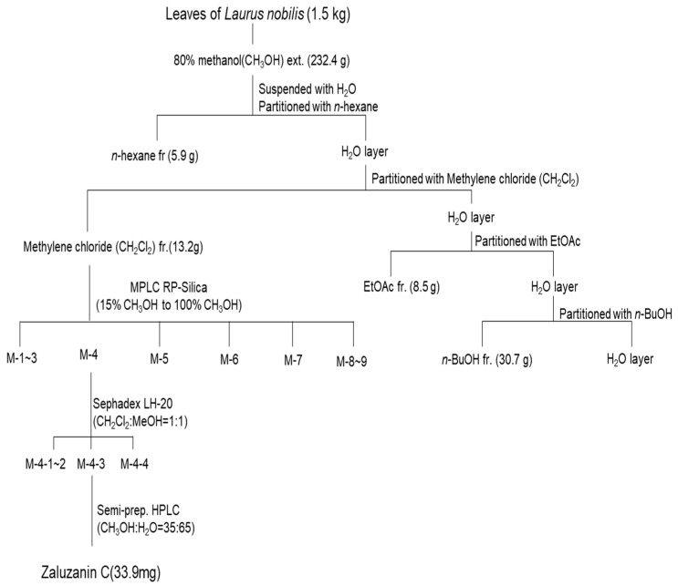
Zaluzanin C (ZC), a sesquiterpene lactone isolated from Laurus nobilis L., has been reported to have anti-inflammatory and antioxidant effects. However, the mechanistic role of ZC in its protective effects in Kupffer cells and hepatocytes has not been elucidated. The purpose of this study was to elucidate the efficacy and mechanism of action of ZC in Kupffer cells and hepatocytes. ZC inhibited LPS-induced mitochondrial ROS (mtROS) production and subsequent mtROS-mediated NF-κB activity in Kupffer cells (KCs). ZC reduced mRNA levels of pro-inflammatory cytokines (Il1b and Tnfa) and chemokines (Ccl2, Ccl3, Ccl4, Cxcl2 and Cxcl9). Tumor necrosis factor (TNF)-α-induced hepatocyte mtROS production was inhibited by ZC. ZC was effective in alleviating mtROS-mediated mitochondrial dysfunction. ZC enhanced mitophagy and increased mRNA levels of fatty acid oxidation genes (Pparα, Cpt1, Acadm and Hadha) and mitochondrial biosynthetic factors (Pgc1α, Tfam, Nrf1 and Nrf2) in hepatocytes. ZC has proven its anti-lipid effect by improving lipid accumulation in hepatocytes by enhancing mitochondrial function to facilitate lipid metabolism. Therefore, our study suggests that ZC may be an effective compound for hepatoprotection by suppressing inflammation and lipid accumulation through regulating mtROS.

Keywords: NAFLD; Zaluzanin C; inflammation; lipid accumulation; mtROS.


Conflict of interest statement

The authors declare no conflict of interest.

Figures

Figure 1
Figure 1
ZC inhibits LPS-induced ROS in Immortalized mouse kupffer cells (ImKC). (A) Structure of ZC. (B) Quantification of ZC cytotoxicity in ImKCs and cells counting assay with 10 μM ZC in ImKCs. Cytotoxicity assay was performed using the Quanti-LDH™ Cytotoxicity Assay Kit. ImKCs were measured using a fluorescence microscope and microplate reader. (C) Representative Western blotting analysis of SOD1. The concentration of LPS was 250 ng/mL, and for ZC, it was 10 μM. (D) MitoSOX 6 h after LPS (500 ng/mL), ZC (10 μM) and Mito-TEMPO (50 μM) treatment. The data are expressed as means ± sem. * p < 0.05, ** p < 0.01.
Figure 2
Figure 2
ZC has anti-inflammatory effects by inhibiting ROS-induced NF-κB signaling. (A) The mRNA expression of cytokines and chemokines treated with LPS (125 ng/mL) and ZC (10 μM) in ImKCs. (B) The mRNA expression of cytokines and chemokines treated with LPS (125 ng/mL), ZC (10 μM) and Mito-TEMPO (50 μM) in ImKCs. (C) Representative Western blotting analysis of p-NF-κB, NF-κB and p-IκB, Actin. The concentration of LPS was 250 ng/mL and ZC was 10 μM and Mito-TEMPO was 50 μM. (D) The protein level of TNF-α in ImKC lysates were determined using Enzyme-linked Immunosorbent Assay. Relative mRNA expression levels were normalized to mouse GAPDH levels. The data are expressed as means ± sem. ns p > 0.05, * p < 0.05, ** p < 0.01, *** p < 0.001, and **** p < 0.0001.
Figure 3
Figure 3
ZC inhibits ROS in hepatocytes induced by TNF-α secreted by Immortalized mouse kupffer cells (ImKCs). (A) Hep3B cells and primary hepatocytes were treated with different ZC concentrations (5 μM to 1000 μM) and cytotoxicity was quantified. (B) Mitochondrial superoxides (MitoSOXs) were measured in Hep3B cells treated with supernatant collected from Immortalized mouse kupffer cells (ImKCs). (C) mRNA expression of TNFR1 after TNFR1 siRNA transfection in primary hepatocytes. (D) Primary hepatocytes transfected with TNFR1 siRNA and scrambled RNA control (SC) were treated with TNF-α (100 ng/mL) for 24 h, and MitoSOXs were measured with a microplate reader. (E) Hep3B cells were measured using a microplate reader 24 h after treatment with TNF-α (100 ng/mL), ZC (10 μM) and Mito-TEMPO (50 μM). (F) Primary hepatocytes were observed by fluorescence microscopy 6 h after treatment with TNF-α (10 ng/mL), ZC (10 μM) and Mito-TEMPO (50 μM). Data are presented as mean ± sem. ns p > 0.05, * p < 0.05, ** p < 0.01, and **** p < 0.0001.
Figure 4
Figure 4
ZC alleviates mitochondrial dysfunction. (A) Mitophagy detection after ZC and Mito-TEMPO treated with for 24 h. ZC- and Mito-TEMPO-mediated mitophagy was quantified by Flow Cytometry with 2 μM carbonyl cyanide m-chlorophenylhydrazone (CCCP). (B) qPCR analysis in primary hepatocytes for mitochondrial DNA (16s) expression normalized to HK2. (CF) mRNA expression of mitochondrial biogenesis factors treated with TNF-a (30 ng/mL) and ZC (10 μM) in Hep3B cells. The data are expressed as means ± sem. * p < 0.05, ** p < 0.01, and **** p < 0.0001.
Figure 5
Figure 5
ZC improves ROS-induced lipid accumulation through β-oxidation activity. The mRNA levels of the beta-oxidation gene markers Acox1 (A), Ucp2 (B), CPT1 (C), ACADM (D) and HADHA (E) were measured by qRT-PCR in Hep3B cells. Hep3B cells were treated with TNF-α 30 ng/mL and ZC 10 μM. (F) BODIPY staining in primary hepatocytes. (G) Quantification of BODIPY/DAPI-positive areas. Relative mRNA expression levels were normalized to human actin levels. Data are presented as mean ± sem. * p < 0.05, ** p < 0.01, *** p < 0.001, and **** p < 0.0001.
Scheme 1
Scheme 1
Isolation method of ZC from leaves of L. nobilis.

Similar articles

Cited by

References

    1. Benedict M., Zhang X. Non-alcoholic fatty liver disease: An expanded review. World J. Hepatol. 2017;9:715–732. doi: 10.4254/wjh.v9.i16.715. - DOI - PMC - PubMed
    1. Byrne C.D., Targher G. NAFLD: A multisystem disease. J. Hepatol. 2015;62((Suppl. S1)):S47–S64. doi: 10.1016/j.jhep.2014.12.012. - DOI - PubMed
    1. Heyens L.J.M., Busschots D., Koek G.H., Robaeys G., Francque S. Liver Fibrosis in Non-alcoholic Fatty Liver Disease: From Liver Biopsy to Non-invasive Biomarkers in Diagnosis and Treatment. Front. Med. 2021;8:615978. doi: 10.3389/fmed.2021.615978. - DOI - PMC - PubMed
    1. Pydyn N., Miękus K., Jura J., Kotlinowski J. New therapeutic strategies in nonalcoholic fatty liver disease: A focus on promising drugs for nonalcoholic steatohepatitis. Pharmacol. Rep. 2020;72:1–12. doi: 10.1007/s43440-019-00020-1. - DOI - PubMed
    1. Chen Y.-Y., Yeh M.M. Non-alcoholic fatty liver disease: A review with clinical and pathological correlation. Pt 1J. Formos. Med. Assoc. 2021;120:68–77. doi: 10.1016/j.jfma.2020.07.006. - DOI - PubMed