. 2023 Nov 9;28(22):7502.
doi: 10.3390/molecules28227502.

Assessing the Protective Role of Epigallocatechin Gallate (EGCG) against Water-Pipe Smoke-Induced Toxicity: A Comparative Study on Gene Expression and Histopathology

Affiliations

Assessing the Protective Role of Epigallocatechin Gallate (EGCG) against Water-Pipe Smoke-Induced Toxicity: A Comparative Study on Gene Expression and Histopathology

Wajdy Al-Awaida et al. Molecules. .

Abstract

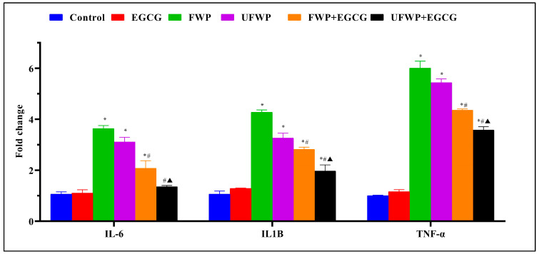
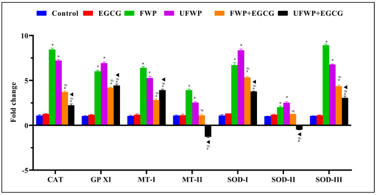
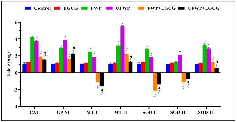
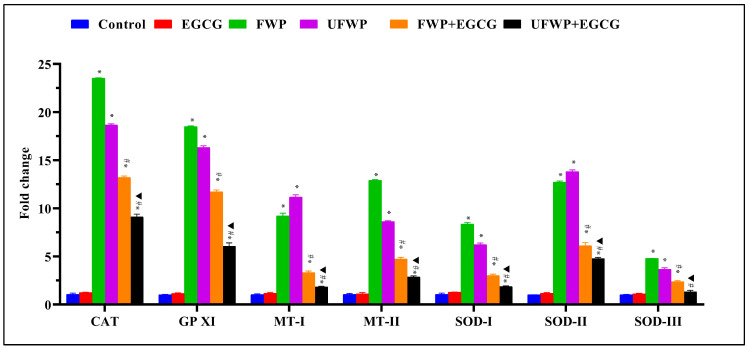
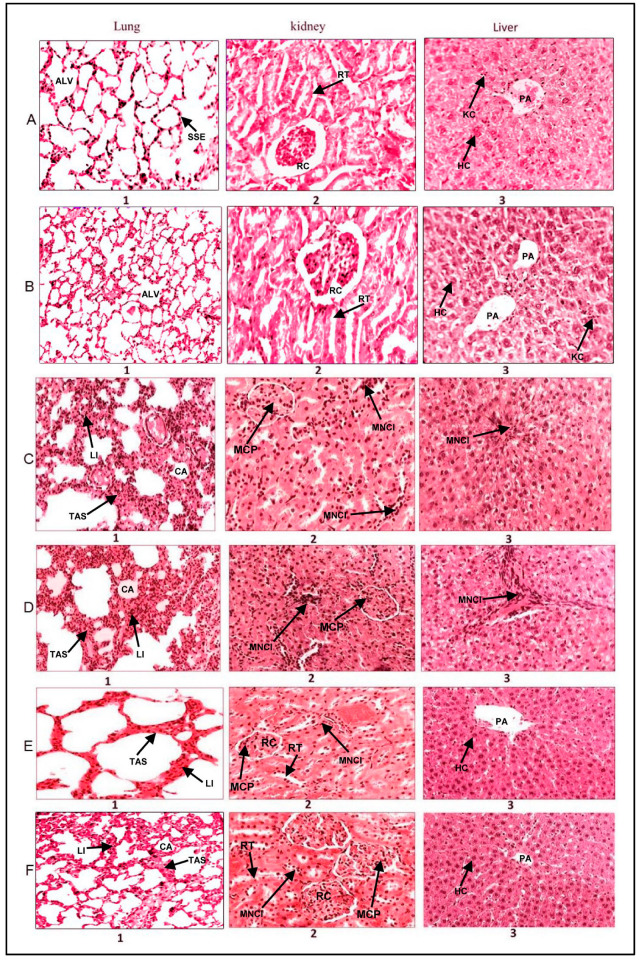
Exposure to water-pipe smoking, whether flavored or unflavored, has been shown to instigate inflammation and oxidative stress in BALB/c mice. This consequently results in alterations in the expression of inflammatory markers and antioxidant genes. This study aimed to scrutinize the impact of Epigallocatechin gallate (EGCG)-a key active component of green tea-on inflammation and oxidative stress in BALB/c mice exposed to water-pipe smoke. The experimental setup included a control group, a flavored water-pipe smoke (FWP) group, an unflavored water-pipe smoke (UFWP) group, and EGCG-treated flavored and unflavored groups (FWP + EGCG and UFWP + EGCG). Expression levels of IL-6, IL1B, TNF-α, CAT, GPXI, MT-I, MT-II, SOD-I, SOD-II, and SOD-III were evaluated in lung, liver, and kidney tissues. Histopathological changes were also assessed. The findings revealed that the EGCG-treated groups manifested a significant decline in the expression of inflammatory markers and antioxidant genes compared to the FWP and UFWP groups. This insinuates that EGCG holds the capacity to alleviate the damaging effects of water-pipe smoke-induced inflammation and oxidative stress. Moreover, enhancements in histopathological features were observed in the EGCG-treated groups, signifying a protective effect against tissue damage induced by water-pipe smoking. These results underscore the potential of EGCG as a protective agent against the adverse effects of water-pipe smoking. By curbing inflammation and oxidative stress, EGCG may aid in the prevention or mitigation of smoking-associated diseases.

Keywords: antioxidant genes; epigallocatechin gallate (EGCG); gene expression; histopathological examination; inflammatory markers; oxidative stress; water-pipe smoke.


Conflict of interest statement

The authors declare no conflict of interest.

Figures

Figure 1
Figure 1
The Anti-Inflammatory Effects of Epigallocatechin Gallate (EGCG) on Inflammatory Markers IL-6, IL1B, and TNF-α in BALB/c mice Exposed to Flavored and Unflavored Water-Pipe Smoke. The figure represents the expression levels of inflammatory markers (IL-6, IL1B, and TNF-α) in control, FWP, UFWP, FWP + EGCG, and UFWP + EGCG groups. Exposure to both flavored and unflavored water-pipe smoke (FWP and UFWP groups) induced upregulation of these markers relative to the control group. However, EGCG treatment effectively reduced this overexpression in the FWP + EGCG and UFWP + EGCG groups, indicating its anti-inflammatory effects. Each value represents the mean ± standard deviation derived from multiple independent experiments. Statistical significance was set at p < 0.05. The keys (*, #, ▲) denote the specific significant differences between groups, *: Represents a significant difference between the control group and all other groups, #: Represents a significant difference between the FWP group and the FWP + EGCG group, ▲: Represents a significant difference between the UFWP group and the UFWP + EGCG group. The threshold for statistical significance was set at p < 0.05.
Figure 2
Figure 2
Modulating Effects of Epigallocatechin Gallate (EGCG) on Antioxidant Gene Expression in Kidney Tissues Exposed to Flavored and Unflavored Water-Pipe Smoke. The figure shows the relative expression of antioxidant genes (CAT, GPXI, MT-I, MT-II, SOD-I, SOD-II, SOD-III) in control, FWP, UFWP, FWP + EGCG, and UFWP + EGCG groups. For all genes examined, exposure to both flavored and unflavored water-pipe smoke (FWP and UFWP groups) led to an increased expression compared to the control. EGCG treatment effectively reduced these elevated levels in the FWP + EGCG and UFWP + EGCG groups, with MT-II and SOD-II in UFWP + EGCG even presenting with a decrease below control levels. Each value represents the mean ± standard deviation derived from multiple independent experiments. The keys (*, #, ▲) denote the specific significant differences between groups, *: Represents a significant difference between the control group and all other groups, #: Represents a significant difference between the FWP group and the FWP + EGCG group, ▲: Represents a significant difference between the UFWP group and the UFWP + EGCG group. The threshold for statistical significance was set at p < 0.05.
Figure 3
Figure 3
Modulating Effects of Epigallocatechin Gallate (EGCG) on Antioxidant Gene Expression in Liver Tissues Exposed to Flavored and Unflavored Water-Pipe Smoke. The figure illustrates the relative expression of antioxidant genes (CAT, GPXI, MT-I, MT-II, SOD-I, SOD-II, SOD-III) in control, FWP, UFWP, FWP + EGCG, and UFWP + EGCG groups. Animals in the FWP and UFWP groups exhibited an increase in the expression of all measured genes compared to the control, with CAT and GPXI levels significantly reduced upon EGCG treatment. Intriguingly, EGCG treatment resulted in MT-I, SOD-I, and SOD-II expression levels in the FWP + EGCG and UFWP + EGCG groups that were lower than the control group, indicating a potential inhibitory effect of EGCG on these genes’ expression upon smoke exposure. For MT-II and SOD-III, while EGCG treatment reduced expression levels, they remained above control in the FWP + EGCG group and decreased significantly in the UFWP + EGCG group. Each value is presented as the mean ± standard deviation derived from multiple independent experiments. The keys (*, #, ▲) denote the specific significant differences between groups, *: Represents a significant difference between the control group and all other groups, #: Represents a significant difference between the FWP group and the FWP + EGCG group, ▲: Represents a significant difference between the UFWP group and the UFWP + EGCG group. The threshold for statistical significance was set at p < 0.05.
Figure 4
Figure 4
Modulating Effects of Epigallocatechin Gallate (EGCG) on Antioxidant Gene Expression in Lung Tissues Exposed to Flavored and Unflavored Water-Pipe Smoke. The figure shows the relative expression of antioxidant genes (CAT, GPXI, MT-I, MT-II, SOD-I, SOD-II, SOD-III) in control, FWP, UFWP, FWP + EGCG, and UFWP + EGCG groups. In the FWP and UFWP groups, exposure to water-pipe smoke led to a significant increase in the expression of all examined genes compared to control. EGCG treatment notably mitigated this increase across all genes, with both FWP + EGCG and UFWP + EGCG groups displaying significantly reduced expression levels compared to their non-EGCG treated counterparts. Each value represents the mean ± standard deviation derived from multiple independent experiments. The keys (*, #, ▲) denote the specific significant differences between groups, *: Represents a significant difference between the control group and all other groups, #: Represents a significant difference between the FWP group and the FWP + EGCG group, ▲: Represents a significant difference between the UFWP group and the UFWP + EGCG group. The threshold for statistical significance was set at p < 0.05.
Figure 5
Figure 5
Effect of Epigallocatechin Gallate (EGCG) Consumption on the Morphology of Lung, Kidney, and Liver Tissues in Mice Exposed to Water-Pipe Smoke (Hematoxylin and Eosin Stain—H & E). Row (A): Displays typical morphology of the liver, lung, and kidney in a mouse exposed to clean air, which serves as the control. Row (B): Presents the liver, lung, and kidney morphology in an EGCG-treated mouse, demonstrating no observable pathological changes. Row (C): Reveals the morphology of liver, lung, and kidney tissues after exposure to unflavored water-pipe smoke. Specifically, C1, C2, and C3 highlight collapsed alveoli and inflammatory cell infiltration in the lungs, mesangial cell proliferation in kidneys, and inflammation in the liver’s portal area. Row (D): Illustrates the morphology of liver, lung, and kidney tissues in mice exposed to flavored water-pipe smoke. D1, D2, and D3 point to collapsed alveoli and inflammatory cell infiltration in the lungs, mesangial cell proliferation in kidneys, and inflammation in the liver’s portal area. Row (E): Shows the morphology of liver, lung, and kidney tissues in mice exposed to unflavored water-pipe smoke but treated with EGCG. E1, E2, and E3 depict lung tissues with diminished alveolar wall thickening, absence of inflammatory cell infiltration, liver tissues devoid of mesangial cell proliferation, and kidney tissues without portal area inflammation. Row (F): Demonstrates the morphology of liver, lung, and kidney tissues in mice exposed to flavored water-pipe smoke but treated with EGCG. F1, F2, and F3 portray lung tissues with diminished alveolar wall thickening, absence of inflammatory cell infiltration, liver tissues free from mesangial cell proliferation, and kidney tissues without portal area inflammation. Tissue Key: Kidney: RT—Renal Tubules, RC—Renal Corpuscle, MCP—Mesangial Cells Proliferation, MNCI—Mononuclear Leukocytes Infiltration. Liver: HC—Hepatocyte Cell, PA—Portal Area, KC—Kupffer Cells, MNCI—Mononuclear Leukocytes Infiltration. Lung: SSE—Simple Squamous Epithelium, Alv—Alveolar Sac, TAS—Thick Alveolar Septum, MNCI—Mononuclear Leukocytes Infiltration, LI—Lymphocyte Infiltration, CA—Collapsed Alveoli.

Similar articles

Cited by

References

    1. Maziak W., Taleb Z.B., Bahelah R., Islam F., Jaber R., Auf R., Salloum R.G. The global epidemiology of waterpipe smoking. Tob. Control. 2015;24:i3–i12. doi: 10.1136/tobaccocontrol-2014-051903. - DOI - PMC - PubMed
    1. Zheng Z., Xie Z., Li D. Discussion of waterpipe tobacco smoking on reddit. Heliyon. 2022;8:e10635. doi: 10.1016/j.heliyon.2022.e10635. - DOI - PMC - PubMed
    1. Al-Awaida W., Najjar H., Shraideh Z. Structural characterization of rat ventricular tissue exposed to the smoke of two types of waterpipe. Iran. J. Basic Med. Sci. 2015;18:942. doi: 10.22038/IJBMS.2015.5456. - DOI - PMC - PubMed
    1. Khwaldeh A., Siyam A.A., Alzbeede A., Farajallah M., Shraideh Z., Badran D. Ameliorative effects of curcumin and caffeic acid against short term exposure of waterpipe tobacco smoking on lung, heart and kidney in mice. Anat. Cell Biol. 2021;54:93–103. doi: 10.5115/acb.20.200. - DOI - PMC - PubMed
    1. Unsal V., Dalkıran T., Çiçek M., Kölükçü E. The role of natural antioxidants against reactive oxygen species produced by cadmium toxicity: A review. Adv. Pharm. Bull. 2020;10:184. doi: 10.34172/apb.2020.023. - DOI - PMC - PubMed

LinkOut - more resources