. 2023 Nov 10;28(22):7528.
doi: 10.3390/molecules28227528.

New Steroidal Selenides as Proapoptotic Factors

Affiliations

New Steroidal Selenides as Proapoptotic Factors

Izabella Jastrzebska et al. Molecules. .

Abstract

Cytostatic and pro-apoptotic effects of selenium steroid derivatives against HeLa cells were determined. The highest cytostatic activity was shown by derivative 4 (GI50 25.0 µM, almost complete growth inhibition after three days of culture, and over 97% of apoptotic and dead cells at 200 µM). The results of our study (cell number measurements, apoptosis profile, relative expression of apoptosis-related APAF1, BID, and mevalonate pathway-involved HMGCR, SQLE, CYP51A1, and PDHB genes, and computational chemistry data) support the hypothesis that tested selenosteroids induce the extrinsic pathway of apoptosis by affecting the cell membrane as cholesterol antimetabolites. An additional mechanism of action is possible through a direct action of derivative 4 to inhibit PDHB expression in a way similar to steroid hormones.

Keywords: HeLa cells; antimetabolites; cell growth inhibition; gene expression; in vitro culture.


Conflict of interest statement

The authors declare no conflict of interest.

Figures

Scheme 1
Scheme 1
Synthesis of selenosteroids 24.
Figure 1
Figure 1
Microscopic observations of HeLa cells after 48 h of culture on a medium with selenosteroids (structure of selenosteroids according to Scheme 1), bar = 20 µm.
Figure 2
Figure 2
Changes in the number of HeLa cells in culture depending on the concentration of used compound after three days of culture (substrate 1; 1,4-androstadiene-3,17-dione—substrate for synthesis of selenosteroid 2, 3, and 4, according to Scheme 1). Data represent medians ± maximum and minimum values. Asterisks indicate statistically significant differences compared to the control (Mann–Whitney U-test, p < 0.05). The number of cells in the control culture corresponds to zero substrate and selenosteroid concentration.
Figure 3
Figure 3
Comparison (means ± SE) of GI50 for substrate (1) and individual selenosteroids (24) based on data from Figure 2. Asterisks indicate values that differ from substrate 1, and hashtags indicate values differ from selenosteroid 4 (Anova and RIR post-hoc test p < 0.05). The differences in GI50 values between selenosteroids 2 and substrate (1) were not statistically significant.
Figure 4
Figure 4
Comparison of IC50/GI50 values of different selenosteroids taking into account the length of the carbon linker between the selenium atom and the steroid core. (A) Structures described in ref. [4]: values given as mean ± SE; lengthening the chain from 2 to 4 carbon atoms resulted in an approximately 3-fold decrease in IC50. (B) Our experimental data: values given as mean ± SE, where elongation of the carbon chain from 1 to 2 atoms resulted in an approximately 2-fold decrease in GI50.
Figure 5
Figure 5
Comparison of the apoptotic profiles of HeLa control cultures and cultures on media with different concentrations of selenosteroid 2 after three days of culture. At the top are examples of histograms of individual cultures obtained as a result of cytometric analysis. At the bottom is the percentage of median data from a series of experiments. Asterisks indicate statistically significant differences concerning the control (Mann–Whitney U-test, p < 0.05).
Figure 6
Figure 6
Comparison of the apoptotic profiles of HeLa control cultures and cultures on media with different concentrations of selenosteroid 3 after three days of culture. At the top are examples of histograms of individual cultures obtained as a result of cytometric analysis. At the bottom is the percentage of median data from a series of experiments. Asterisks indicate statistically significant differences concerning control (Mann–Whitney U-test, p < 0.05).
Figure 7
Figure 7
Comparison of the apoptotic profiles of HeLa control cultures and on media with different concentrations of selenosteroid 4 after three days of culture. At the top are examples of histograms of individual cultures obtained as a result of cytometric analysis. At the bottom is the percentage of median data from a series of experiments. Asterisks indicate statistically significant differences concerning control (Mann–Whitney U-test, p < 0.05).
Figure 8
Figure 8
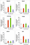
Comparison of the relative expression of selected genes after three days of HeLa cell culture in media supplemented with different selenosteroids (2—red color, 3—green color, 4—purple color) at a concentration equal to the GI50 value as shown in Figure 3. Values are shown as % of control ± standard deviation. Asterisks indicate statistically significant differences compared to the control (Student’s t-test, p < 0.05).
Figure 9
Figure 9
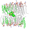
Position of selenosteroid 4 (surface view) in the lipid bilayer model of HeLa lineage cells. Cholesterol molecules forming the layers are highlighted in green. The water molecules and ions were removed for the sake of clarity.
Figure 10
Figure 10
Analysis of RMSD and Rg of unliganded HeLa bilayer model (UNL) and three complexes during 10 ns of molecular dynamics simulation: (a) root mean square deviation (RMSD) for non-hydrogen atoms; (b) radius of gyration (Rg).
Figure 11
Figure 11
Proposed mechanism of action of the tested selenosteroids 24. All tested compounds exhibit cytostatic activity with varying intensity depending on their chemical structure. The proapoptotic effect of selenosteroids may result from the destabilization of the cell membrane and activation of the extrinsic apoptotic pathway related to caspase 8. Elevated expression of BID activated by caspase 8 may enhance apoptosis by releasing cytochrome C from mitochondria. The most cytostatic derivative, 4, additionally reduces PDHB expression, probably through a hormonal pathway, which leads to the disorganization of cell bioenergetics and inhibition of the mevalonate pathway by reducing acetyl-CoA synthesis.
Figure 12
Figure 12
The alignment of the built-in cholesterol moiety from ref. [43] (green scaffold) superimposed on the best pose from the redocking procedure (magenta).

Similar articles

References

    1. Jastrzebska I., Grzes P.A., Niemirowicz-Laskowska K., Car H. Selenosteroids—Promising hybrid compounds with pleiotropic biological activity: Synthesis and biological aspects. J. Steroid Biochem. Mol. Biol. 2021;213:105975. doi: 10.1016/j.jsbmb.2021.105975. - DOI - PubMed
    1. Romero-Hernández L.L., Merino-Montiel P., Montiel-Smith S., Meza-Reyes S., Vega-Báez J.L., Abasolo I., Schwartz S., López Ó., Fernández-Bolaños J.G. Diosgenin-based thio(seleno)ureas and triazolyl glycoconjugates as hybrid drugs. Antioxidant and antiproliferative profile. Eur. J. Med. Chem. 2015;99:67–81. doi: 10.1016/j.ejmech.2015.05.018. - DOI - PubMed
    1. Fuentes-Aguilar A., Romero-Hernández L.L., Arenas-González A., Merino-Montiel P., Montiel-Smith S., Meza-Reyes S., Vega-Báez J.L., Plata G.B., Padrón J.M., López Ó., et al. New selenosteroids as antiproliferative agents. Org. Biomol. Chem. 2017;15:5041–5054. doi: 10.1039/C7OB00458C. - DOI - PubMed
    1. Huang Y., Peng Z., Wei M., Gan C., Zhang Y., Chen S., Xiao J., Cui J. Synthesis and antiproliferative evaluation of some novel estradiol selenocyanates. Steroids. 2022;181:108992. doi: 10.1016/j.steroids.2022.108992. - DOI - PubMed
    1. Huang Y., Peng Z., Wei M., Pang L., Cheng Y., Xiao J.-A., Gan C., Cui J. Straightforward synthesis of steroidal selenocyanates through oxidative umpolung selenocyanation of steroids and their antitumor activity. J. Steroid Biochem. Mol. Biol. 2023;225:106203. doi: 10.1016/j.jsbmb.2022.106203. - DOI - PubMed

LinkOut - more resources