. 2023 Nov 15;28(22):7611.
doi: 10.3390/molecules28227611.

Galactooligosaccharide Mediates NF-κB Pathway to Improve Intestinal Barrier Function and Intestinal Microbiota

Affiliations

Galactooligosaccharide Mediates NF-κB Pathway to Improve Intestinal Barrier Function and Intestinal Microbiota

Menglu Xi et al. Molecules. .

Abstract

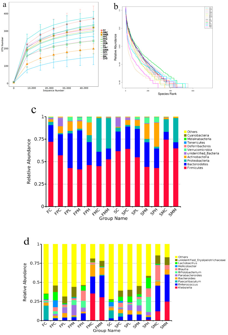
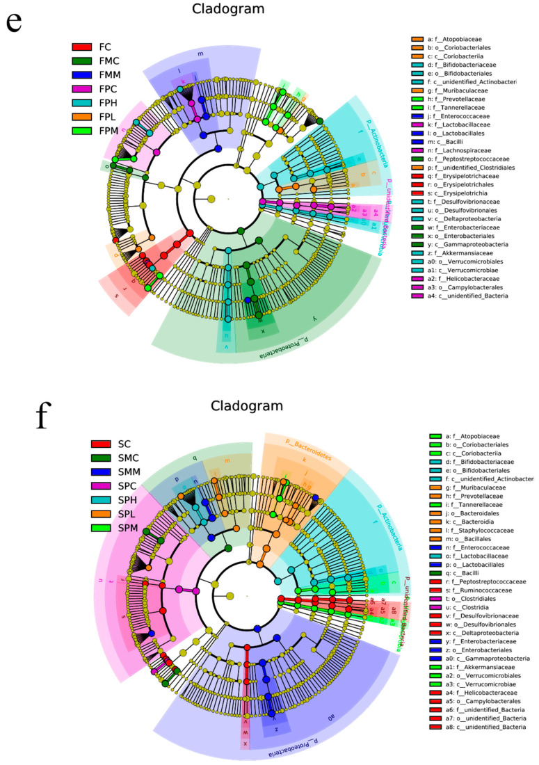
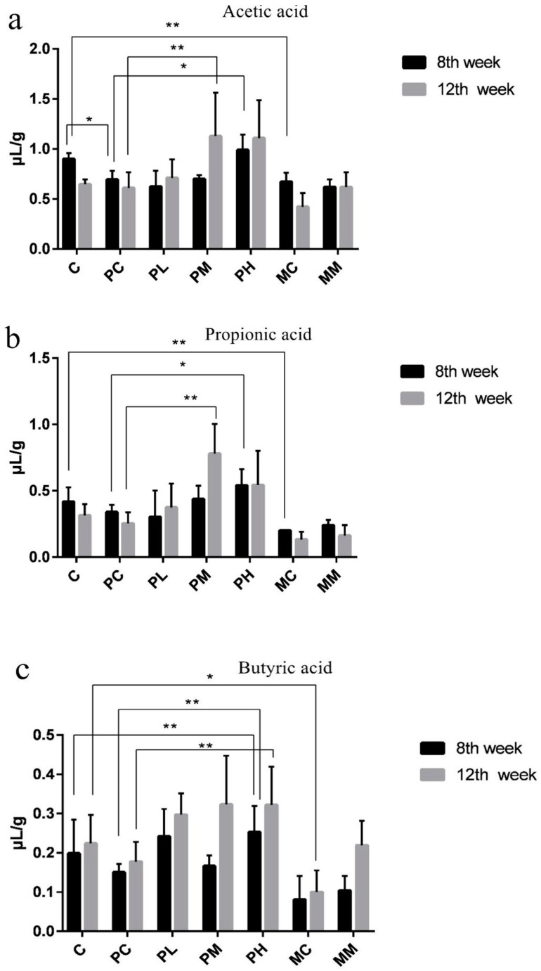
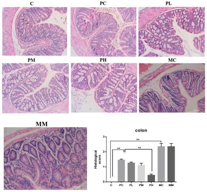
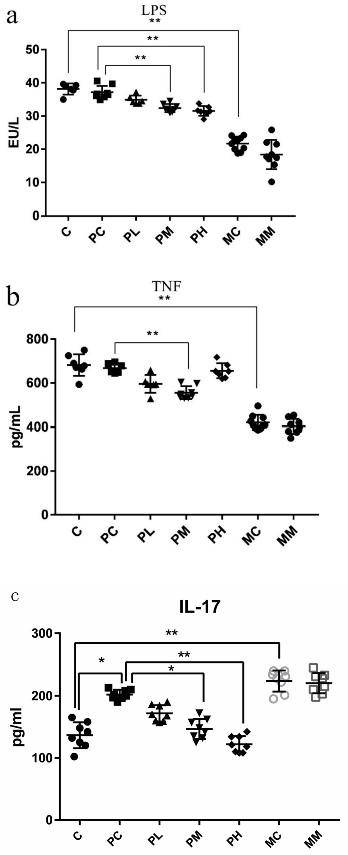
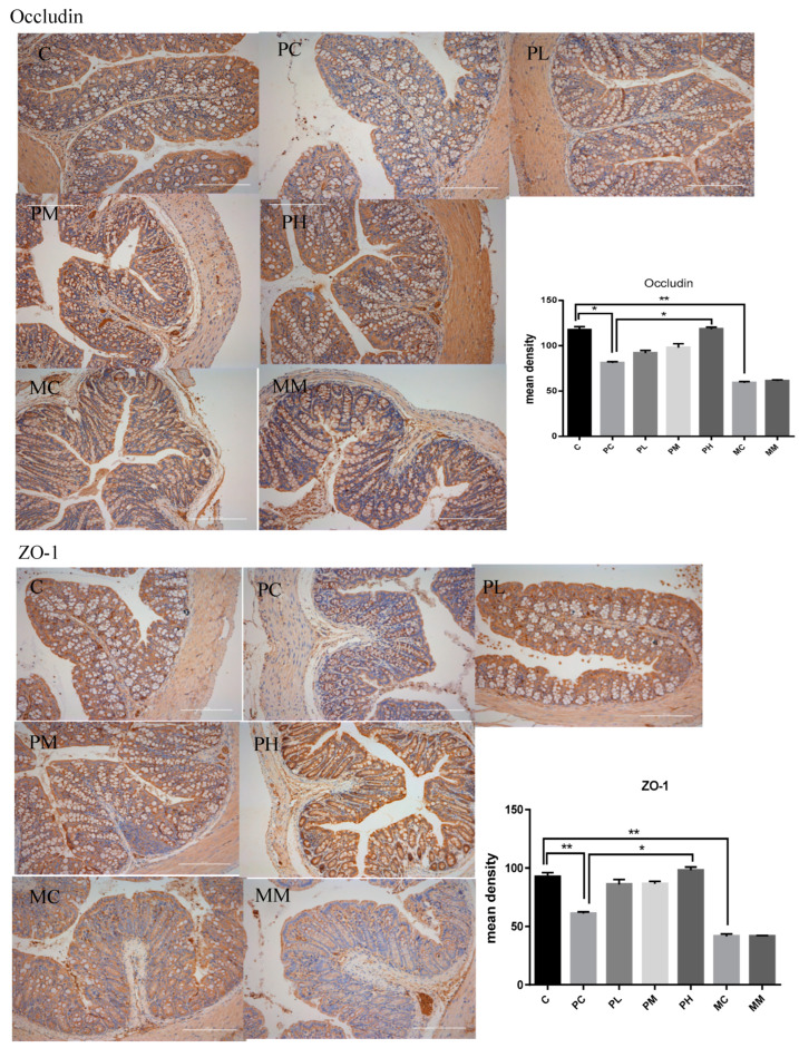
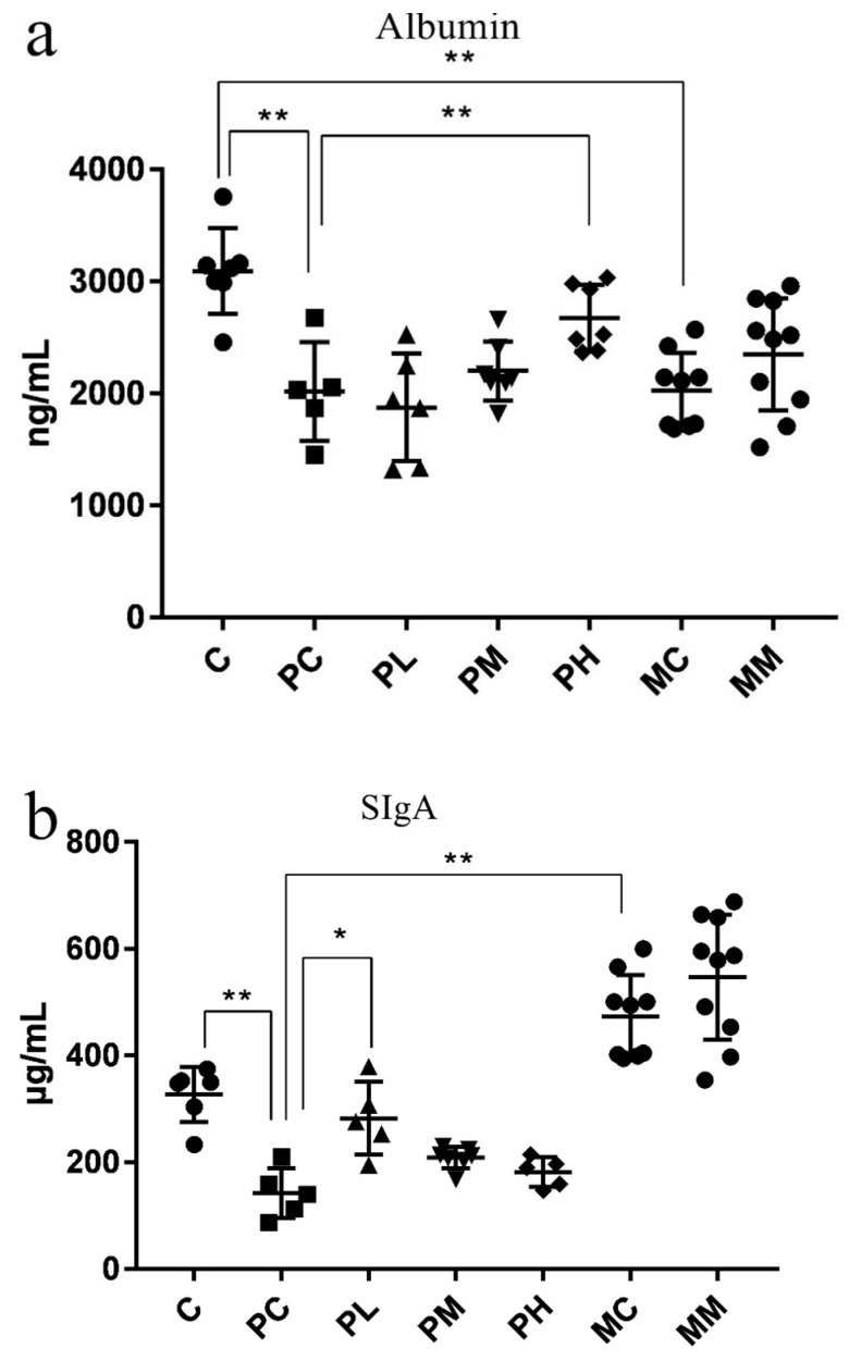
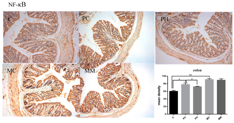
The use of antibiotics to treat diarrhea and other diseases early in life can lead to intestinal disorders in infants, which can cause a range of immune-related diseases. Intestinal microbiota diversity is closely related to dietary intake, with many oligosaccharides impacting intestinal microorganism structures and communities. Thus, oligosaccharide type and quantity are important for intestinal microbiota construction. Galactooligosaccharides (GOS) are functional oligosaccharides that can be supplemented with infant formula. Currently, information on GOS and its impact on intestinal microbiota diversity and disorders is lacking. Similarly, GOS is rarely reported within the context of intestinal barrier function. In this study, 16S rRNA sequencing, gas chromatography, and immunohistochemistry were used to investigate the effects of GOS on the intestinal microbiota and barrier pathways in antibiotic-treated mouse models. The results found that GOS promoted Bifidobacterium and Akkermansia proliferation, increased short-chain fatty acid levels, increased tight junction protein expression (occludin and ZO-1), increased secretory immunoglobulin A (SIgA) and albumin levels, significantly downregulated NF-κB expression, and reduced lipopolysaccharide (LPS), interleukin-IL-1β (IL-1β), and IL-6 levels. Also, a high GOS dose in ampicillin-supplemented animals provided resistance to intestinal damage.

Keywords: galactooligosaccharides; gut microbiota; intestinal barrier; short-chain fatty acids.


Conflict of interest statement

The authors declare no conflict of interest.

Figures

Figure 1
Figure 1
Intestinal microbiota composition after GOS supplementation with ampicillin exposition. (a) Rarefaction curve; (b) Rank curves; (c) Relative contribution of the top 10 phyla in each group; (d) Relative contribution of the top 10 genera in each group; (e,f) Taxonomic cladogram from LEfSe at 8 and 12 weeks, (n = 4). Biomarker taxa are highlighted with colored circles and shaded areas. Circle diameters reflect taxa abundance in the community. A cut-off value of ≥4.0, used for linear discriminant analysis (LDA), is shown. F 8 weeks, S 12 weeks.
Figure 1
Figure 1
Intestinal microbiota composition after GOS supplementation with ampicillin exposition. (a) Rarefaction curve; (b) Rank curves; (c) Relative contribution of the top 10 phyla in each group; (d) Relative contribution of the top 10 genera in each group; (e,f) Taxonomic cladogram from LEfSe at 8 and 12 weeks, (n = 4). Biomarker taxa are highlighted with colored circles and shaded areas. Circle diameters reflect taxa abundance in the community. A cut-off value of ≥4.0, used for linear discriminant analysis (LDA), is shown. F 8 weeks, S 12 weeks.
Figure 2
Figure 2
Short-chain fatty acid levels in feces. Acetic (a), propionic (b), and butyric acid (c) levels in cecal contents were measured using gas chromatography at 8 weeks (black) and 12 weeks (light gray) after study commencement (n = 8). Data (mean ± standard deviation (SD)) were analyzed using one-way ANOVA (* p < 0.05; ** p < 0.01). Data are represented by three biological replicates. C is a normal AIN-93G diet and water; PC is a normal AIN-93G diet and water containing ampicillin; PL is a low-dose GOS AIN-93G diet (0.5% w/w) and water containing ampicillin; PM is a medium-dose GOS AIN-93G diet (2% w/w) and water containing ampicillin; PH is a high-dose GOS AIN-93G diet (5% w/w) and water containing ampicillin; MC is a regular AIN-93G diet and water containing streptomycin (1 g/L), ampicillin (1 g/L), and gentamicin (1 g/L); MM is a medium-dose GOS AIN-93G diet (2% w/w) and water containing streptomycin (1 g/L), ampicillin (1 g/L), gentamicin (1 g/L).
Figure 3
Figure 3
Representative images of hematoxylin and eosin staining (magnification 200×; scale bar = 200 μm); the gray bar chart presents the histological scores of the colon sections (** p < 0.01). C is a normal AIN-93G diet and water; PC is a normal AIN-93G diet and water containing ampicillin; PL is a low-dose GOS AIN-93G diet (0.5% w/w) and water containing ampicillin; PM is a medium-dose GOS AIN-93G diet (2% w/w) and water containing ampicillin; PH is a high-dose GOS AIN-93G diet (5% w/w) and water containing ampicillin; MC is a regular AIN-93G diet and water containing streptomycin (1 g/L), ampicillin (1 g/L), and gentamicin (1 g/L); MM is a medium-dose GOS AIN-93G diet (2% w/w) and water containing streptomycin (1 g/L), ampicillin (1 g/L), gentamicin (1 g/L).
Figure 4
Figure 4
Assessing inflammation, immunocompetence, and intestinal permeability biomarkers using enzyme-linked immunosorbent assay. In (ac), the concentrations of LPS, TNF-α, and IL-17 in mouse serum are represented, respectively (n = 8). Data (mean ± standard deviation (SD)) were analyzed using one-way ANOVA (* p < 0.05; ** p < 0.01).
Figure 5
Figure 5
Immunohistochemistry of occludin and ZO-1 in the colon. The bottom panel presents the mean density of immunohistochemistry. Data (mean ± SD) were analyzed with one-way ANOVA (* p < 0.05; ** p < 0.01).
Figure 6
Figure 6
(a,b) Evaluating intestinal barrier immunity and tight junctions by measuring SIgA and fecal albumin levels (n = 8). Data (mean ± standard deviation (SD)) were analyzed using one-way ANOVA (* p < 0.05; ** p < 0.01).
Figure 7
Figure 7
Immunohistochemistry of NF-κB in the colon. The bottom panel presents the mean density of immunohistochemistry. Data (mean ± SD) were analyzed with one-way ANOVA (* p < 0.05; ** p < 0.01).

Similar articles

Cited by

References

    1. Sun Y., Song J., Lan X., Ma F., Jiang M., Jiang C. Calcium-Sensitive Receptors Alters Intestinal Microbiota Metabolites Especially SCFAs and Ameliorates Intestinal Barrier Damage in Neonatal Rat Endotoxemia. Infect. Drug. Resist. 2023;16:5707–5717. doi: 10.2147/IDR.S420689. - DOI - PMC - PubMed
    1. Zhang L.Z., Gong J.G., Li J.H., Hao Y.S., Xu H.J., Liu Y.C., Feng Z.H. Dietary resveratrol supplementation on growth performance, immune function and intestinal barrier function in broilers challenged with lipopolysaccharide. Poultry Sci. 2023;102:102968. doi: 10.1016/j.psj.2023.102968. - DOI - PMC - PubMed
    1. Castro M., Valero M.S., López-Tofiño Y., López-Gómez L., Girón R., Martín-Fontelles M.I., Uranga J.A., Abalo R. Radiographic and histopathological study of gastrointestinal dysmotility in lipopolysaccharide-induced sepsis in the rat. Neurogastroent. Motil. 2023;35:e14639. doi: 10.1111/nmo.14639. - DOI - PubMed
    1. Sommer F., Bäckhed F. The gut microbiota—Masters of host development and physiology. Nat. Rev. Microbiol. 2013;11:227–238. doi: 10.1038/nrmicro2974. - DOI - PubMed
    1. Yoo J.S., Oh S.F. Unconventional immune cells in the gut mucosal barrier: Regulation by symbiotic microbiota. Exp. Mol. Med. 2023;55:1905–1912. doi: 10.1038/s12276-023-01088-9. - DOI - PMC - PubMed

LinkOut - more resources