. 2023 Nov 15;23(22):9191.
doi: 10.3390/s23229191.

Optofluidic Flow Cytometer with In-Plane Spherical Mirror for Signal Enhancement

Affiliations

Optofluidic Flow Cytometer with In-Plane Spherical Mirror for Signal Enhancement

Filippo Zorzi et al. Sensors (Basel). .

Abstract

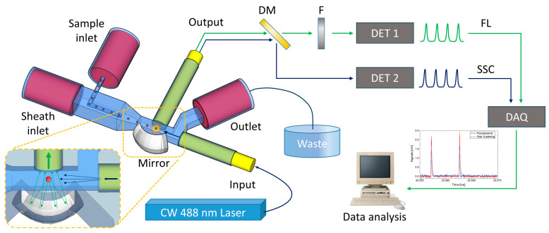
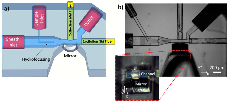
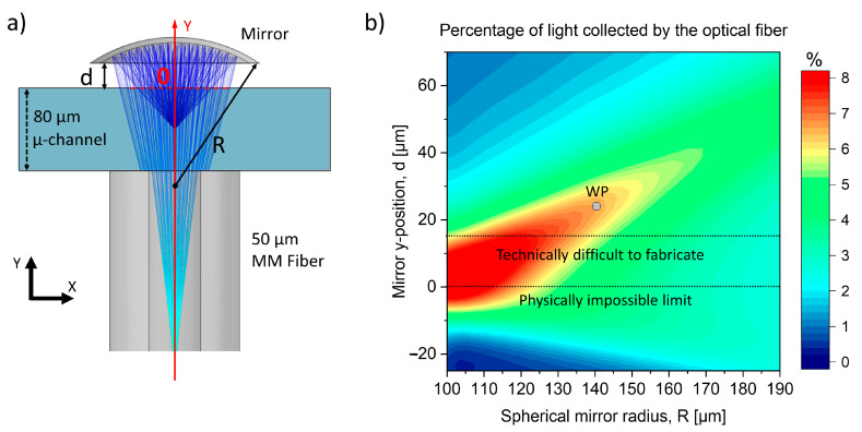
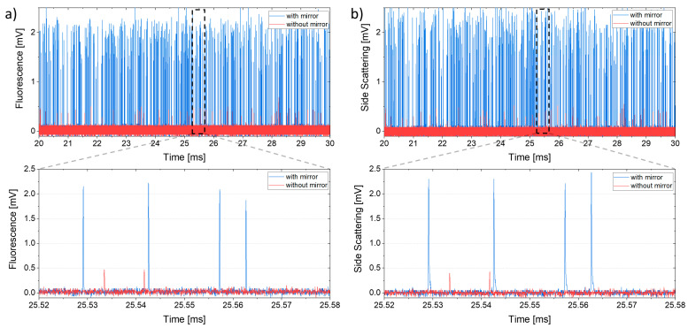
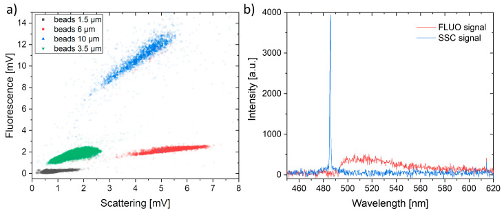
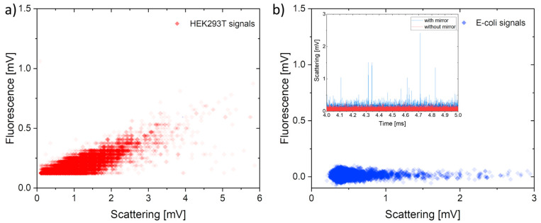
Statistical analysis of the properties of single microparticles, such as cells, bacteria or plastic slivers, has attracted increasing interest in recent years. In this regard, field flow cytometry is considered the gold standard technique, but commercially available instruments are bulky, expensive, and not suitable for use in point-of-care (PoC) testing. Microfluidic flow cytometers, on the other hand, are small, cheap and can be used for on-site analyses. However, in order to detect small particles, they require complex geometries and the aid of external optical components. To overcome these limitations, here, we present an opto-fluidic flow cytometer with an integrated 3D in-plane spherical mirror for enhanced optical signal collection. As a result, the signal-to-noise ratio is increased by a factor of six, enabling the detection of particle sizes down to 1.5 µm. The proposed optofluidic detection scheme enables the simultaneous collection of particle fluorescence and scattering using a single optical fiber, which is crucial to easily distinguishing particle populations with different optical properties. The devices have been fully characterized using fluorescent polystyrene beads of different sizes. As a proof of concept for potential real-world applications, signals from fluorescent HEK cells and Escherichia coli bacteria were analyzed.

Keywords: FLICE; Lab on a Chip; femtosecond laser microfabrication; flow cytometry; optofluidic particles detection.


Conflict of interest statement

The authors declare no conflict of interest.

Figures

Figure 1
Figure 1
Sketch of the optofluidic chip and the detection apparatus. Once placed in the center of the outer channel via hydrodynamic focusing, the sample is excited, and the emitted light is collected by an optical fiber. The signal is divided into two spectral regions by a dichroic mirror (DM). A notch filter (F) was used in order to cut the pump beam in the fluorescent branch. The signals are converted by two photodetectors (DET 1 and 2), acquired by a DAQ and finally processed by NI LabVIEW 2017 software that provides a real-time analysis of the collected data.
Figure 2
Figure 2
(a) Sketch of the optofluidic chip geometry. (b) Bright-field microscope image of the fabricated chip acquired with 5× objective lens. The image was acquired during the hydrodynamic focusing tests performed by pumping isopropanol inside the focusing channel and water in the sheath inlet. Zoom—side view of the mirror before the metallization.
Figure 3
Figure 3
(a) Sketch of the geometry used for the simulation made with COMSOL Multiphysics. (b) Results obtained from the simulations. The working point (WP) corresponds to the mirror with a radius of 140 µm and with its center located at 24 µm from the microchannel.
Figure 4
Figure 4
Comparison of fluorescence (a) and side scattering (b) signals from 3.5 µm beads obtained with (blue) and without (red) the integration of the spherical mirror.
Figure 5
Figure 5
(a) Scattering and fluorescence signals obtained using a sample with a mix of fluorescent polystyrene spheres. (b) Spectral analysis of the light collected by the fibers after the signal splitting.
Figure 6
Figure 6
(a) Correlation between scattering and fluorescence signals obtained by analyzing HEK293T cells made fluorescent by CellMaskTM molecules. (b) Signals obtained by analyzing a water sample contaminated by Escherichia coli bacteria using the chip with a spherical mirror. In this case, the fluorescence signal was below the detection limit. Incept—time behavior of the E. coli scattering signal obtained with (blue) and without (red) the spherical mirror.

Similar articles

Cited by

References

    1. Rangel J.M., Sparling P.H., Crowe C., Griffin P.M., Swerdlow D.L. Epidemiology of Escherichia Coli O157:H7 Outbreaks, United States, 1982–2002. Emerg. Infect. Dis. 2005;11:603–609. doi: 10.3201/eid1104.040739. - DOI - PMC - PubMed
    1. World Health Organization. UNICEF . Ending Preventable Child Deaths from Pneumonia and Diarrhoea by 2025: The Integrated Global Action Plan for Pneumonia and Diarrhoea (GAPPD) World Health Organization; Geneva, Switzerland: 2013.
    1. Ankeny J.S., Court C.M., Hou S., Li Q., Song M., Wu D., Chen J.F., Lee T., Lin M., Sho S., et al. Circulating Tumour Cells as a Biomarker for Diagnosis and Staging in Pancreatic Cancer. Br. J. Cancer. 2016;114:1367–1375. doi: 10.1038/bjc.2016.121. - DOI - PMC - PubMed
    1. Leng S.X., McElhaney J.E., Walston J.D., Xie D., Fedarko N.S., Kuchel G.A. ELISA and Multiplex Technologies for Cytokine Measurement in Inflammation and Aging Research. J. Gerontol. A Biol. Sci. Med. Sci. 2008;63:879–884. doi: 10.1093/gerona/63.8.879. - DOI - PMC - PubMed
    1. Yang S.-Y., Lien K.-Y., Huang K.-J., Lei H.-Y., Lee G.-B. Micro Flow Cytometry Utilizing a Magnetic Bead-Based Immunoassay for Rapid Virus Detection. Biosens. Bioelectron. 2008;24:855–862. doi: 10.1016/j.bios.2008.07.019. - DOI - PubMed

MeSH terms