

Macrophage-Derived Factors with the Potential to Contribute to Pathogenicity of HIV-1 and HIV-2: Role of CCL-2/MCP-1
- PMID: 38005838
- PMCID: PMC10674259
- DOI: 10.3390/v15112160
Macrophage-Derived Factors with the Potential to Contribute to Pathogenicity of HIV-1 and HIV-2: Role of CCL-2/MCP-1
Abstract
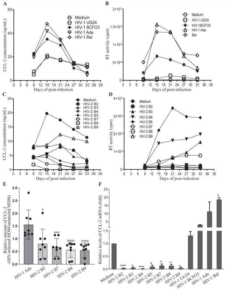
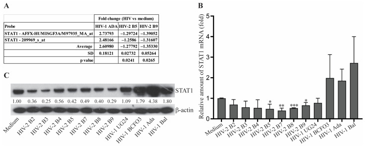
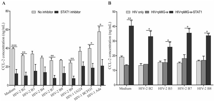
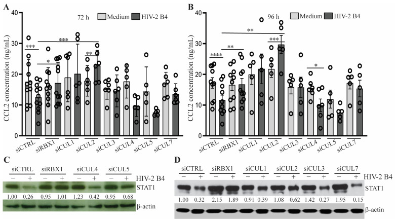
Human immunodeficiency virus type 2 (HIV-2) is known to be less pathogenic than HIV-1. However, the mechanism(s) underlying the decreased HIV-2 pathogenicity is not fully understood. Herein, we report that β-chemokine CCL2 expression was increased in HIV-1-infected human monocyte-derived macrophages (MDM) but decreased in HIV-2-infected MDM when compared to uninfected MDM. Inhibition of CCL2 expression following HIV-2 infection occurred at both protein and mRNA levels. By microarray analysis, quantitative PCR, and Western blotting, we identified that Signal Transducer and Activator of Transcription 1 (STAT1), a critical transcription factor for inducing CCL2 gene expression, was also reduced in HIV-2-infected MDM. Blockade of STAT1 in HIV-infected MDM using a STAT1 inhibitor significantly reduced the production of CCL2. In contrast, transduction of STAT1-expressing pseudo-retrovirus restored CCL2 production in HIV-2-infected MDM. These findings support the concept that CCL2 inhibition in HIV-2-infected MDM is meditated by reduction of STAT1. Furthermore, we showed that STAT1 reduction in HIV-2-infected MDM was regulated by the CUL2/RBX1 ubiquitin E3 ligase complex-dependent proteasome pathway. Knockdown of CUL2 or RBX1 restored the expression of STAT1 and CCL2 in HIV-2-infected MDM. Taken together, our findings suggest that differential regulation of the STAT1-CCL2 axis may be one of the mechanisms underlying the different pathogenicity observed for HIV-1 and HIV-2.
Keywords: CCL2; CULLIN 2; HIV-1; HIV-2; RBX1; STAT1; macrophage.
Conflict of interest statement
The authors declare that they have no conflict of interest with the contents of this article.
Figures
Similar articles
-
Reversible Human Immunodeficiency Virus Type-1 Latency in Primary Human Monocyte-Derived Macrophages Induced by Sustained M1 Polarization.Sci Rep. 2018 Sep 24;8(1):14249. doi: 10.1038/s41598-018-32451-w. Sci Rep. 2018. PMID: 30250078 Free PMC article.
-
Transcriptome Profiling of Human Monocyte-Derived Macrophages Upon CCL2 Neutralization Reveals an Association Between Activation of Innate Immune Pathways and Restriction of HIV-1 Gene Expression.Front Immunol. 2020 Sep 18;11:2129. doi: 10.3389/fimmu.2020.02129. eCollection 2020. Front Immunol. 2020. PMID: 33072075 Free PMC article.
-
Human immunodeficiency virus replication induces monocyte chemotactic protein-1 in human macrophages and U937 promonocytic cells.Blood. 1999 Mar 15;93(6):1851-7. Blood. 1999. PMID: 10068657
-
Regulation of human immunodeficiency virus type 1 infection, beta-chemokine production, and CCR5 expression in CD40L-stimulated macrophages: immune control of viral entry.J Virol. 2001 May;75(9):4308-20. doi: 10.1128/JVI.75.9.4308-4320.2001. J Virol. 2001. PMID: 11287580 Free PMC article.
-
The macrophage response to HIV-1: Intracellular control of X4 virus replication accompanied by activation of chemokine and cytokine synthesis.J Neurovirol. 2002 Dec;8(6):599-610. doi: 10.1080/13550280290100923. J Neurovirol. 2002. PMID: 12476353 Review.
Cited by
-
Roles of Macrophages in Viral Infections.Viruses. 2024 Oct 21;16(10):1643. doi: 10.3390/v16101643. Viruses. 2024. PMID: 39459975 Free PMC article.
-
Macrophage-Derived Factors with the Potential to Contribute to the Pathogenicity of HIV-1 and HIV-2: Roles of M-CSF and CXCL7.Int J Mol Sci. 2025 May 23;26(11):5028. doi: 10.3390/ijms26115028. Int J Mol Sci. 2025. PMID: 40507836 Free PMC article.
References
-
- Kedzierska K., Crowe S.M. The role of monocytes and macrophages in the pathogenesis of HIV-1 infection. Curr. Med. Chem. 2002;9:1893–1903. - PubMed
-
- Reeves J.D., Doms R.W. Human immunodeficiency virus type 2. Pt 6J. Gen. Virol. 2002;83:1253–1265. - PubMed
-
- Freedman D., Shattock A., Stuart J., McLaughlin H. Acquired immunodeficiency syndrome. Ir. Med. J. 1989;82:135–138. - PubMed
-
- de Silva T.I., Cotten M., Rowland-Jones S.L. HIV-2: The forgotten AIDS virus. Trends Microbiol. 2008;16:588–595. - PubMed
-
- Berry N., Ariyoshi K., Jaffar S., Sabally S., Corrah T., Tedder R., Whittle H. Low peripheral blood viral HIV-2 RNA in individuals with high CD4 percentage differentiates HIV-2 from HIV-1 infection. J. Hum. Virol. 1998;1:457–468. - PubMed
MeSH terms
Substances
Grants and funding
LinkOut - more resources
Full Text Sources
Medical
Research Materials
Miscellaneous
