. 2023 Oct 27;15(11):2161.
doi: 10.3390/v15112161.

Chemoprophylactic Assessment of Combined Intranasal SARS-CoV-2 Polymerase and Exonuclease Inhibition in Syrian Golden Hamsters

Affiliations

Chemoprophylactic Assessment of Combined Intranasal SARS-CoV-2 Polymerase and Exonuclease Inhibition in Syrian Golden Hamsters

Eduardo Gallardo-Toledo et al. Viruses. .

Abstract

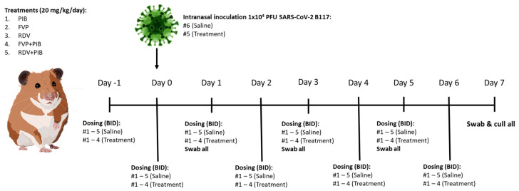
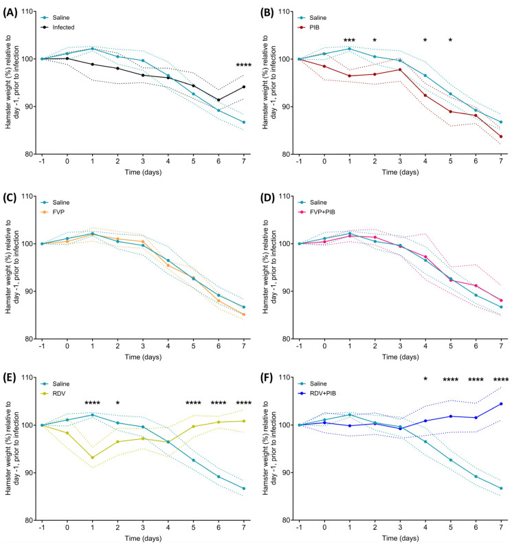
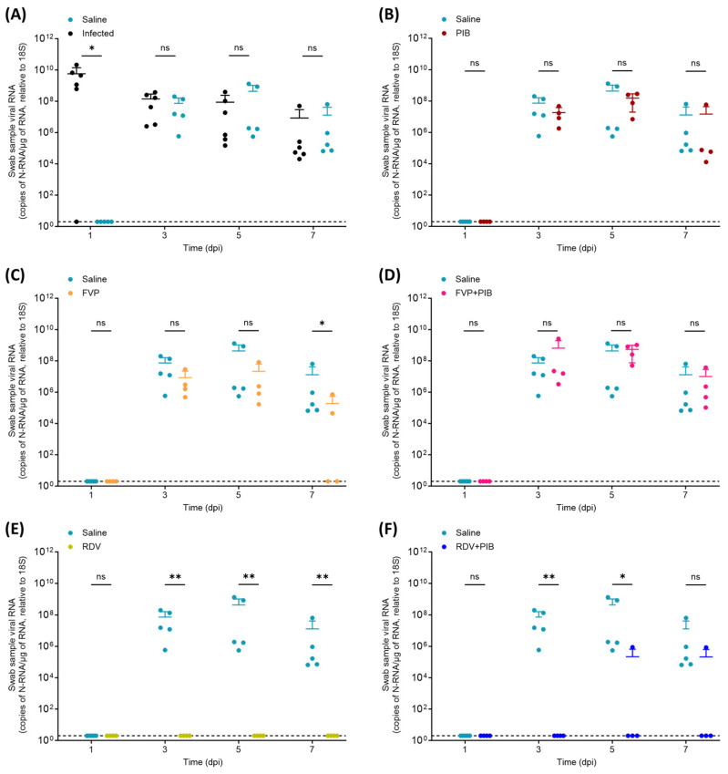
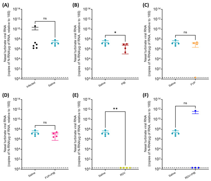
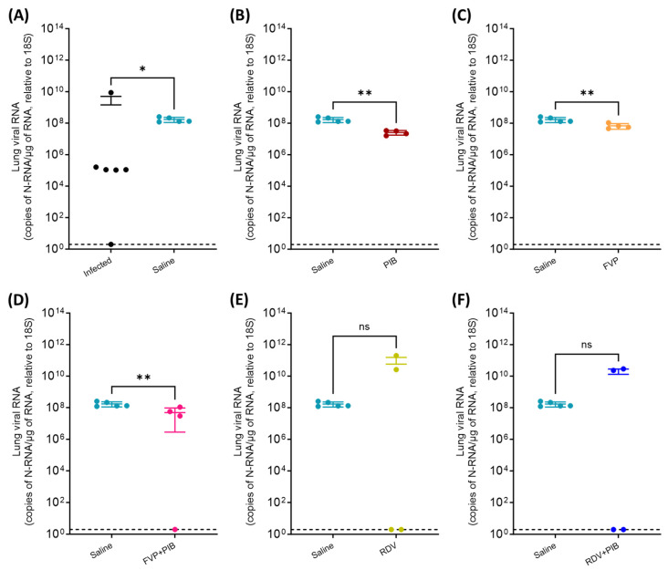
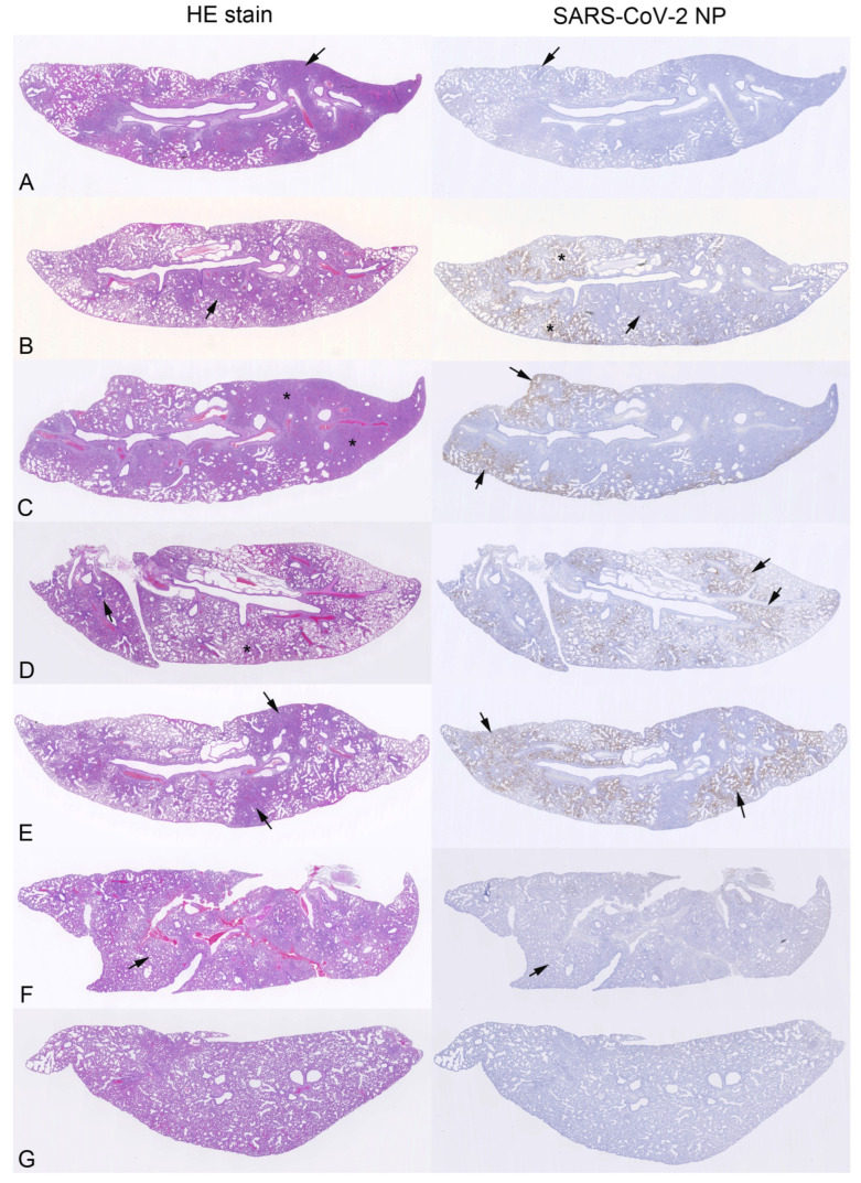
Pibrentasvir (PIB) has been demonstrated to block exonuclease activity of the SARS-CoV-2 polymerase, protecting favipiravir (FVP) and remdesivir (RDV) from post-incorporation excision and eliciting antiviral synergy in vitro. The present study investigated the chemoprophylactic efficacy of PIB, FVP, RDV, FVP with PIB, or RDV with PIB dosed intranasally twice a day, using a Syrian golden hamster contact transmission model. Compared to the saline control, viral RNA levels were significantly lower in throat swabs in FVP (day 7), RDV (day 3, 5, 7), and RDV+PIB (day 3, 5) treatment groups. Similarly, findings were evident for nasal turbinate after PIB and RDV treatment, and lungs after PIB, FVP, and FVP+PIB treatment at day 7. Lung viral RNA levels after RDV and RDV+PIB treatment were only detectable in two animals per group, but the overall difference was not statistically significant. In situ examination of the lungs confirmed SARS-CoV-2 infection in all animals, except for one in each of the RDV and RDV+PIB treatment groups, which tested negative in all virus detection approaches. Overall, prevention of transmission was observed in most animals treated with RDV, while other agents reduced the viral load following contact transmission. No benefit of combining FVP or RDV with PIB was observed.

Keywords: SARS-CoV-2; chemoprophylaxis; favipiravir; pibrentasvir; remdesivir.


Conflict of interest statement

A.O. is a director of Tandem Nano Ltd. and co-inventor of drug delivery patents. A.O. has been co-investigator on funding received by the University of Liverpool from ViiV Healthcare and Gilead Sciences in the past 3 years, unrelated to COVID-19. A.O. has received personal fees from Gilead and Assembly Biosciences in the past 3 years, also unrelated to COVID-19. J.P.S. has received funding from Byotrol Technologies, ENA Respiratory, Sisaf, and Zucero not related to the current abstract.

Figures

Figure 1
Figure 1
Study design for evaluation of the chemoprophylactic efficacy of pibrentasvir (PIB), favipiravir (FVP), remdesivir (RDV), FVP+PIB, or RDV+PIB dosed intranasally twice a day (BID); 10 mg/kg BID, total 20 mg/kg/day. The control group was dosed with saline. Treatments started 24 h prior to being co-housed with an untreated hamster intranasally inoculated with 1 × 10⁴ PFU SARS-CoV-2 B117. All animals were throat swabbed at day 1, 3, 5, and 7.
Figure 2
Figure 2
Hamsters from each group (n = 4, except saline n = 5) and infected hamsters (n = 6) were weighed daily from day −1 to day 7 post infection. All weights are shown as a percentage of the initial weight recorded at baseline, day −1 of the study. (A) Saline vs. infected, (B) Saline vs. PIB, (C) Saline vs. FVP, (D) Saline vs. FVP with PIB, (E) Saline vs. RDV, (F) Saline vs. RDV with PIB. Statistical significance was determined by 2-way ANOVA multiple comparison with Bonferroni correction. * = p ≤ 0.05, *** = p ≤ 0.001, **** = p ≤ 0.0001. Dotted lines represent standard deviations.
Figure 3
Figure 3
Quantification of SARS-CoV-2 N-RNA in throat swabs taken on day 1, 3, 5, and 7 post intranasal inoculation of the donor (i.e., infected) hamsters. (A) Saline vs. Infected, (B) Saline vs. PIB, (C) Saline vs. FVP, (D) Saline vs. FVP+PIB, (E) Saline vs. RDV, (F) Saline vs. RDV+PIB. To determine statistical significance (p ≤ 0.05), a nonparametric Mann–Whitney test (one-tailed) was performed between the saline control treated group (n = 5) and the infected group (n = 6) or the corresponding chemoprophylaxis-treated group (n = 4) for each day. ns = not statistically significant, * = p ≤ 0.05, ** = p ≤ 0.01. LOD: limit of detection (indicated by dotted line).
Figure 4
Figure 4
Quantification of SARS-CoV-2 N-RNA in nasal turbinate samples at day 7 post intranasal inoculation of the donor (i.e., infected) hamsters. (A) Saline vs. Infected, (B) Saline vs. PIB, (C) Saline vs. FVP, (D) Saline vs. FVP+PIB, (E) Saline vs. RDV, (F) Saline vs. RDV+PIB. Comparison between saline control group (n = 5) and the infected group (n = 6) or the corresponding chemoprophylaxis-treated group (n = 4) was performed to determine statistical significance (nonparametric Mann–Whitney test [one-tailed], p ≤ 0.05). ns = not statistically significant, * = p ≤ 0.05, ** = p ≤ 0.01. LOD: limit of detection (indicated by dotted line).
Figure 5
Figure 5
Quantification of SARS-CoV-2 N-RNA in lung samples at day 7 post intranasal inoculation of the donor (i.e., infected) hamsters. Comparison between saline control group (n = 5) and treatment groups (n = 4). (A) Saline vs. Infected, (B) Saline vs. PIB, (C) Saline vs. FVP, (D) Saline vs. FVP+PIB, (E) Saline vs. RDV, (F) Saline vs. RDV+PIB. Statistical significance (p ≤ 0.05) was determined using a nonparametric Mann–Whitney test (one-tailed) between the saline control group (n = 5) and the infected group (n = 6) or the corresponding chemoprophylaxis-treated group (n = 4). ns = not statistically significant, * = p ≤ 0.05, ** = p ≤ 0.01. LOD: limit of detection (indicated by dotted line).
Figure 6
Figure 6
Histological changes and viral antigen expression in the lungs of hamsters intranasally infected with SARS-CoV-2 (A) or treated with saline (B), PIB (C), FVP (D), FVP with PIB (E), RDV (F), or RDV with PIB (G) and housed together as a group of 5 hamsters with an infected hamster; animals were euthanised and examined at day 7. Left column: HE-stained sections; right column: consecutive sections stain for SARS-CoV-2 nucleoprotein (NP), immunohistology, and haematoxylin counterstain. (A) Infected hamster (#3). The lung parenchyma exhibits multifocal-to-coalescing consolidated areas (arrow). Viral antigen expression (right image) is seen in alveolar epithelial cells (AECs) in consolidated areas and patches of unaltered alveoli. Arrows: area shown in higher magnification in Supplementary Figure S1A. (B) Saline-treated control animal (#4). The histological changes are dominated by multifocal areas of alveoli that contain desquamed AEC and/or alveolar macrophages (AMs) and exhibit activated type II pneumocytes, some syncytial cells and some degenerate cells. There is widespread viral antigen expression in AEC in large patches of alveoli (*) and, less extensively, in consolidated areas (arrow). Arrows: area shown in higher magnification in Supplementary Figure S1B. (C) PIB-treated hamster (#4). The lung parenchyma exhibits focal extensive consolidation (*). There is widespread viral antigen expression in AEC in large patches of alveoli (arrows). (D) FVP-treated hamster (#2). The lung parenchyma exhibits focal desquamative changes (*) and small focal consolidated areas with epithelial hyperplasia (arrow). There is widespread viral antigen expression in AEC in large patches of alveoli (arrows, right image). (E) FVP+PIB-treated animal (#2). The lung parenchyma exhibits focal consolidated areas with focal epithelial hyperplasia (arrows, left image). There is widespread viral antigen expression in AEC in large patches of alveoli (arrows, right image). (F) RDV-treated hamster (#1). The lung parenchyma appears cell rich (due to increased interstitial cellularity), but otherwise unaltered. There is no evidence of viral antigen expression (right image). Arrows: area shown in higher magnification in Supplementary Figure S1C. (G) RDV-with-PIB-treated hamster (#4). The lung parenchyma is widely unaltered, and there is no evidence of viral antigen expression.

Similar articles

Cited by

References

    1. Dhama K., Nainu F., Frediansyah A., Yatoo M.I., Mohapatra R.K., Chakraborty S., Zhou H., Islam M.R., Mamada S.S., Kusuma H.I., et al. Global emerging Omicron variant of SARS-CoV-2: Impacts, challenges and strategies. J. Infect. Public Health. 2023;16:4–14. doi: 10.1016/j.jiph.2022.11.024. - DOI - PMC - PubMed
    1. Meng Z., Ma D., Duan S., Zhang J., Yue R., Li X., Gao Y., Zeng F., Xu X., Jiang G., et al. Immunological Study of Combined Administration of SARS-CoV-2 DNA Vaccine and Inactivated Vaccine. Vaccines. 2022;10:929. doi: 10.3390/vaccines10060929. - DOI - PMC - PubMed
    1. Wang Z., Yang L. The Therapeutic Potential of Natural Dietary Flavonoids against SARS-CoV-2 Infection. Nutrients. 2023;15:3443. doi: 10.3390/nu15153443. - DOI - PMC - PubMed
    1. Debnath S.K., Debnath M., Srivastava R., Omri A. Drugs repurposing for SARS-CoV-2: New insight of COVID-19 druggability. Expert Rev. Anti Infect. Ther. 2022;20:1187–1204. doi: 10.1080/14787210.2022.2082944. - DOI - PubMed
    1. Qi H., Liu B., Wang X., Zhang L. The humoral response and antibodies against SARS-CoV-2 infection. Nat. Immunol. 2022;23:1008–1020. doi: 10.1038/s41590-022-01248-5. - DOI - PubMed