. 2023 Oct 31;15(11):2202.
doi: 10.3390/v15112202.

Crystal Structure of Inhibitor-Bound GII.4 Sydney 2012 Norovirus 3C-Like Protease

Affiliations

Crystal Structure of Inhibitor-Bound GII.4 Sydney 2012 Norovirus 3C-Like Protease

Alice-Roza Eruera et al. Viruses. .

Abstract

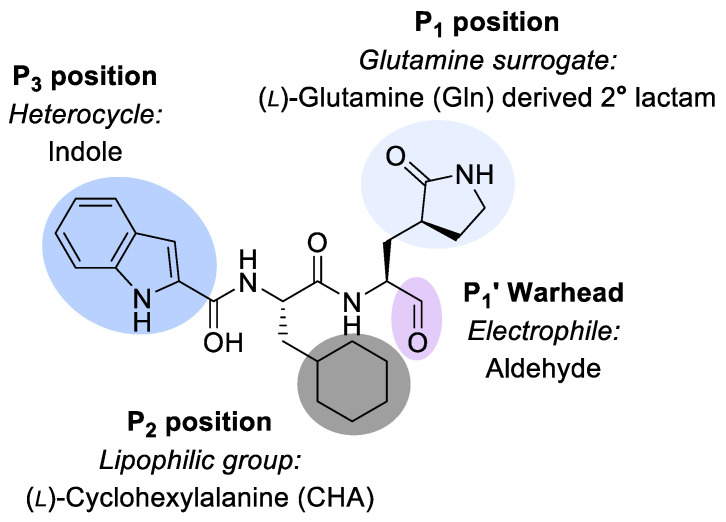
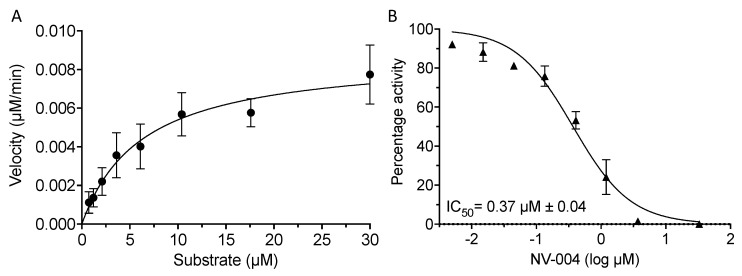
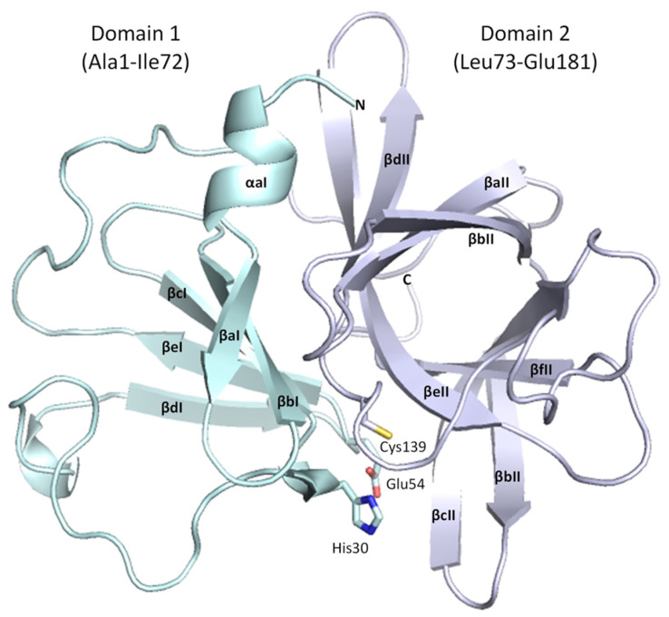
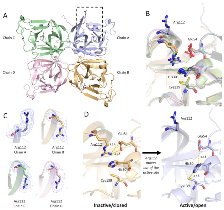
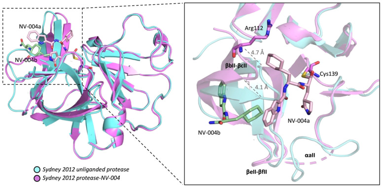
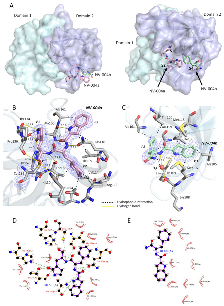
Norovirus is the leading cause of viral gastroenteritis worldwide, and there are no approved vaccines or therapeutic treatments for chronic or severe norovirus infections. The structural characterisation of the norovirus protease and drug development has predominantly focused upon GI.1 noroviruses, despite most global outbreaks being caused by GII.4 noroviruses. Here, we determined the crystal structures of the GII.4 Sydney 2012 ligand-free norovirus protease at 2.79 Å and at 1.83 Å with a covalently bound high-affinity (IC50 = 0.37 µM) protease inhibitor (NV-004). We show that the active sites of the ligand-free protease structure are present in both open and closed conformations, as determined by their Arg112 side chain orientation. A comparative analysis of the ligand-free and ligand-bound protease structures reveals significant structural differences in the active site cleft and substrate-binding pockets when an inhibitor is covalently bound. We also report a second molecule of NV-004 non-covalently bound within the S4 substrate binding pocket via hydrophobic contacts and a water-mediated hydrogen bond. These new insights can guide structure-aided drug design against the GII.4 genogroup of noroviruses.

Keywords: 3C-like protease; X-ray structure; antiviral; ligand-free protease; norovirus; protease inhibitor.


Conflict of interest statement

The authors declare no conflict of interest. The funders had no role in the design of the study; in the collection, analyses, or interpretation of data; in the writing of the manuscript; or in the decision to publish the results.

Figures

Figure 1
Figure 1
NV-004 structure. NV-004 contains an aldehyde as its electrophilic warhead (P1′, lilac), as well as a glutamine analogue (light grey, P1), a lipophilic group (dark grey, P2), and an indole heterocyclic group (blue, P3).
Figure 2
Figure 2
The synthetic scheme for NV-004 production. Reagents and conditions: (a) (i) 4 M HCl/1,4-dioxane, 0 °C to room temperature (rt), 1 h; (ii) Boc-Cha-OH, HCTU, NMM, DMF, 0 °C, 1 h, 86%; (b) (i) 4 M HCl/1,4-dioxane, 0 °C to rt, 1 h; (ii) indole-2-carboxylic acid, HCTU, NMM, DMF, 0 °C, 1.5 h; (c) NaBH4, MeOH, rt, 6.5 h, 63% over 2 steps; (d) Dess–Martin periodinane, CH2Cl2, 0 °C to rt, 2 h, 47%.
Figure 3
Figure 3
Kinetics and dose response curve of NV-004 with GII.4 Sydney 2012 Pro. (A) GII.4 Pro proteins (0.5 μM) were incubated with substrate and the reaction velocity as a function of substrate concentration (0.73–30 μM) was plotted. Kinetic values of 6.3 ± 3.3 μM, 2.9 × 10⁻⁴ ± 0.3 × 10⁻⁴ s−1, and 46.4 M−1 s−1 were determined for the Km, kcat, and kcat/Km, respectively. (B) GII.4 Pro at 0.5 μM was incubated with NV-004 for 40 min, following which, FRET peptide substrate (4 μM) was added. Normalised protease activity was plotted against log inhibitor concentration and the IC50 (μM) was calculated with GraphPad Prism software. Data represent the mean and standard deviation of three biological repeats.
Figure 4
Figure 4
Overall structure of GII.4 HuNoV Sydney 2012 protease. Domain I is in cyan, and Domain II is in mauve. Alpha helices and beta strands are labelled consistent with the standardised nomenclature for norovirus proteases. This figure depicts Chain B from the ligand-free GII.4 HuNoV Sydney 2012 protease structure in PyMOL.
Figure 5
Figure 5
Conformation of Arg112 affects the active site residues. (A) The asymmetric unit of GII.4 HuNoV protease (ligand-free) is shown as a protein cartoon. Chain A is purple, Chain B is orange, Chain C is green, Chain D is pink. Residues His30, Glu54, Arg112, and Cys139 are shown as sticks. The active site and Arg112 are indicated in a dashed box for Chain A with the active site open. (B) Overlay of the four chains in the ASU, showing a distinct conformation for Arg112 in Chain A. (C) Polder maps (3.0 σ) for Arg112 in Chains A–D. (D) Changes in active site conformation between Chain B (inactive/closed) and Chain A (active/open). Polar interactions between Arg112 and active site residues His30, Glu54, and Cys139 are indicated with dashed yellow lines.
Figure 6
Figure 6
GII.4 HuNoV Sydney 2012 protease with bound inhibitor NV-004. Protease-NV-004 is shown in ribbon representation (hot pink) superposed on the unliganded protease (blue). NV-004a (pink, site 1) in the active site and NV-004b (green, site 2) in the S4 pocket are displayed in stick representation. Distances were measured in USCF ChimeraX. NV-004 molecules are shown as sticks with oxygen atoms in red and nitrogen atoms in blue. Active site Cys139 side chain shown as a stick. Structural elements are labelled in bold.
Figure 7
Figure 7
NV-004 bound to GII.4 HuNoV Sydney 2012 protease. (A) Location of NV-004 bound to GII.4 HuNoV Protease. The protease is shown in two different orientations. In each orientation, one protomer of the protease is displayed as a surface representation coloured by the domain. Domain I is in cyan; Domain II is in purple. The two NV-004 molecules are shown as sticks. NV-004a (pink) is located at the active site (S1–S2), and NV-004b (green) is in a nearby site (S4). Subpockets are labelled in bold. (B) Polder map density (contoured to 3σ) for NV-004a is shown as a blue mesh. NV-004a (pink sticks) is in the S1/S2 subsite. Residues of protease that interact with NV-004a are shown as sticks (grey). Hydrogen bonds are shown as solid yellow lines. Hydrophobic interactions are shown as dashed black lines. (C). Polder map density (contoured to 3σ) for NV-004b is shown as a blue mesh. NV-004b (green sticks) is found in the S4 subsite. Residues of protease that interact with NV-004b are shown as sticks (grey). A water molecule that forms a water-mediated H-bond with NV-004b and the protease is shown as a red sphere. (D). LigPLOT diagram for NV-004a. (E) LigPLOT diagram for NV-004b displaying the bonds involved in the interactions between the NV-004 and GII.4 HuNoV protease. Green lines denote hydrogen bonds, with distances shown in Å; the thin purple line denotes the covalent bond between NV-004 and the protein. Red radials represent non-bonded hydrophobic interactions.

Similar articles

Cited by

References

    1. Banyai K., Estes M.K., Martella V., Parashar U.D. Viral gastroenteritis. Lancet. 2018;392:175–186. doi: 10.1016/S0140-6736(18)31128-0. - DOI - PMC - PubMed
    1. Koo H.L., Neill F.H., Estes M.K., Munoz F.M., Cameron A., DuPont H.L., Atmar R.L. Noroviruses: The Most Common Pediatric Viral Enteric Pathogen at a Large University Hospital After Introduction of Rotavirus Vaccination. J. Pediatric Infect. Dis. Soc. 2013;2:57–60. doi: 10.1093/jpids/pis070. - DOI - PMC - PubMed
    1. Lopman B.A., Steele D., Kirkwood C.D., Parashar U.D. The Vast and Varied Global Burden of Norovirus: Prospects for Prevention and Control. PLoS Med. 2016;13:e1001999. doi: 10.1371/journal.pmed.1001999. - DOI - PMC - PubMed
    1. Siebenga J.J., Vennema H., Zheng D.P., Vinje J., Lee B.E., Pang X.L., Ho E.C., Lim W., Choudekar A., Broor S., et al. Norovirus illness is a global problem: Emergence and spread of norovirus GII.4 variants, 2001–2007. J. Infect. Dis. 2009;200:802–812. doi: 10.1086/605127. - DOI - PubMed
    1. Winder N., Gohar S., Muthana M. Norovirus: An Overview of Virology and Preventative Measures. Viruses. 2022;14:2811. doi: 10.3390/v14122811. - DOI - PMC - PubMed