. 2023 Nov 3;15(11):2211.
doi: 10.3390/v15112211.

Human Complement Inhibits Myophages against Pseudomonas aeruginosa

Affiliations

Human Complement Inhibits Myophages against Pseudomonas aeruginosa

Julia E Egido et al. Viruses. .

Abstract

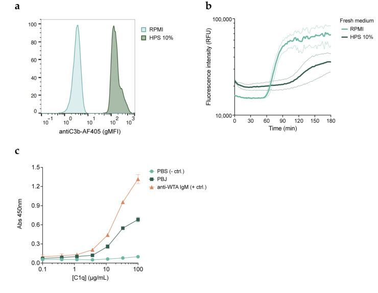
Therapeutic bacteriophages (phages) are primarily chosen based on their in vitro bacteriolytic activity. Although anti-phage antibodies are known to inhibit phage infection, the influence of other immune system components is less well known. An important anti-bacterial and anti-viral innate immune system that may interact with phages is the complement system, a cascade of proteases that recognizes and targets invading microorganisms. In this research, we aimed to study the effects of serum components such as complement on the infectivity of different phages targeting Pseudomonas aeruginosa. We used a fluorescence-based assay to monitor the killing of P. aeruginosa by phages of different morphotypes in the presence of human serum. Our results reveal that several myophages are inhibited by serum in a concentration-dependent way, while the activity of four podophages and one siphophage tested in this study is not affected by serum. By using specific nanobodies blocking different components of the complement cascade, we showed that activation of the classical complement pathway is a driver of phage inhibition. To determine the mechanism of inhibition, we produced bioorthogonally labeled fluorescent phages to study their binding by means of microscopy and flow cytometry. We show that phage adsorption is hampered in the presence of active complement. Our results indicate that interactions with complement may affect the in vivo activity of therapeutically administered phages. A better understanding of this phenomenon is essential to optimize the design and application of therapeutic phage cocktails.

Keywords: Pseudomonas aeruginosa; complement system; phage therapy.


Conflict of interest statement

The authors declare no conflict of interest. The funders had no role in the design of the study; in the collection, analyses, or interpretation of data; in the writing of the manuscript; or in the decision to publish the results.

Figures

Figure 1
Figure 1
Human serum inhibits lytic activity of myophages. Pseudomonas aeruginosa strain PAO1 was incubated with bacteriophages (phages) PBJ, 14-1, LKD16, or LUZ19 at 37 °C in the presence of the DNA dye SYTOX Green. Phage-mediated damage is signaled by an increase in the fluorescence intensity of SYTOX Green. (a) Fluorescence intensity (relative fluorescence units, RFUs) over time of PAO1 infected with different phages at a multiplicity of infection (MOI) of 1 in the absence of serum. Uninfected bacteria were used as the control. (b) Fluorescence intensity (RFUs) over time of PAO1 infected with different phages at an MOI of 1 in the presence of 10% human pooled serum (HPS). The control is uninfected bacteria incubated with 10% HPS. (cf) Time of damage induction (min) by phage (c) PBJ, (d) 14-1, (e) LDK16, and (f) LUZ19, at a range of MOIs (0.1, 1, 10) combined with HPS at different concentrations (0–30%). Time of damage induction is determined as the time after the addition of phages at which the fluorescence curve experiences a sharp and steady increase (1000 RFUs over the previous measurement). (af) Data represent mean ± SD of three independent experiments.
Figure 2
Figure 2
Human serum inhibits a variety of myophages targeting PAO1. (a) Time of damage induction (min) of the different phages as determined by the fluorescent DNA dye assay. Time of damage induction is determined here as the time after the addition of phages at which the fluorescence curve experiences a sharp and steady increase (700 RFUs over the previous measurement). Phages were added at an MOI of 1 and incubated with PAO1 at 37 °C in the absence of serum (control) or in the presence of 10% HPS. (b) S. aureus strain ATCC 19685 was incubated with phage K at an MOI of 10 in the absence (control) or presence of 10% HPS for 2 h at 37 °C, after which bacteria were plated and incubated overnight. Number of recovered colonies per plated volume is expressed as colony forming units per mL (CFUs/mL). Black dotted line represents detection limit. (a,b) Data represent mean ± SD of three independent experiments.
Figure 3
Figure 3
Inhibition of phages by serum is mediated by the complement system. PAO1 was incubated with phage PBJ at 37 °C in the presence of the DNA dye SYTOX Green. (a) Fluorescence intensity (RFUs) over time (min) of bacteria infected with PBJ at an MOI of 10 (control), in the presence of 10% HPS, 10% HPS with heat-inactivated complement (HI HPS), or IgG and IgM purified from HPS at a concentration equivalent to 10%. (b) Fluorescence intensity (RFUs) over time (min) of bacteria infected with PBJ at an MOI of 10 (control), in the presence of 10% HPS, 10% HPS with 50 μM compstatin, or 10% HPS with 20 μg/mL OmCI. (c) Fluorescence intensity (RFUs) of bacteria after 90 min of infection with PBJ at an MOI of 10 in the presence of 10% HPS and nanobodies at a range of concentrations. Value for bacteria infected with phage in the absence of serum is shown as a control. (d) Fluorescence intensity (RFUs) over time (min) of bacteria infected with PBJ at an MOI of 10 in the absence of serum (control) or in the presence of 10% HPS and nanobodies at a concentration of 3 μM. (ad) Data represent mean ± SD of three independent experiments.
Figure 4
Figure 4
Phages tagged with an azide group and labeled with a fluorescent marker were used to study their binding to bacteria. (a) Schematic view of the technique used to produce azido-tagged phages. A methionine auxotroph PAO1 mutant was cultured in minimal medium supplemented with L-azidohomoalanine. Phages were amplified overnight at 37 °C using methionine auxotroph PAO1 as the host. The resulting phage progeny incorporates the non-canonical amino acid L-azidohomoalanine in place of methionine in its proteins, making them available for biorthogonal labeling via click chemistry. (b,c) PAO1-sfCherry (red) was incubated with PBJ-AF488 (green, MOI 50) for 5 min at 37 °C in (b) buffer or (c) 10% HPS, fixed in 1% PFA, washed, and immobilized on agar pads. Images were acquired with a 100× immersion objective and are overlays of the modes phase contrast and widefield fluorescence with cube filters. Images are representative of three independent experiments. (dg) Flow cytometry was performed on PAO1-sfCherry incubated with PBJ-AF488 at different MOIs. When indicated, 10% HPS, anti-C1q nanobody (1 μM), or anti-C3 nanobody (1 μM) was added for 5 min at 37 °C. Samples were stained with monoclonal anti-C3-AF405 antibody and fixed in 1% PFA before measuring. Bacteria were gated on sfCherry-height and forward scatter-height signals. (d) Geometric mean of the signal corresponding to bound phages. Heat-inactivated (HI) phage was used as a negative control. (e) Histograms of phage fluorescent signal at an MOI of 200 or HI phage at an MOI of 400 in the various serum conditions. (f) Histograms of fluorescent signal corresponding to C3b deposition (AF405) on bacteria treated with the various serum conditions. (g) Geometric mean of the signal corresponding to C3b (AF405) for the conditions with PBJ-AF488 at an MOI of 200, or HI phage at an MOI of 400. Statistical analysis was performed using an ordinary one-way ANOVA with Tukey’s multiple comparisons test. Significance is shown as ** p ≤ 0.01 or *** p ≤ 0.001. (d,g) Data represent mean ± SD of three independent experiments. (e,f) Signal was normalized by number of events. A representative graph of three independent experiments is shown.
Figure 5
Figure 5
Inhibition of phages by the complement system is not caused by blockage of phage receptors. (a) Flow cytometry histograms showing C3b deposition (AF405) on bacteria incubated with 10% HPS for 30 min at 37 °C after removal of unbound serum components and staining with aC3b-AF405 nanobody. Signal was normalized by number of events. A representative graph of three independent experiments is shown. (b) Fluorescence intensity (RFUs) over time of complement pre-opsonized PAO1 incubated at 37 °C with phage PBJ (MOI 10) added either in buffer (RPMI) or in 10% HPS in the presence of SYTOX Green. Data represent mean ± SD of three independent experiments. (c) An ELISA was performed to assess the binding of C1q to phage PBJ. Wells coated with buffer were used as a negative control, while wells coated with an anti-S. aureus WTA human IgM were used as a positive control. Data represent mean ± SD of an experiment performed in triplicate.

Similar articles

Cited by

References

    1. Gordillo Altamirano F.L., Barr J.J. Phage Therapy in the Postantibiotic Era. Clin. Microbiol. Rev. 2019;32:e00066-18. doi: 10.1128/CMR.00066-18. - DOI - PMC - PubMed
    1. Strathdee S.A., Hatfull G.F., Mutalik V.K., Schooley R.T. Phage Therapy: From Biological Mechanisms to Future Directions. Cell. 2023;186:17–31. doi: 10.1016/j.cell.2022.11.017. - DOI - PMC - PubMed
    1. Hatfull G.F., Dedrick R.M., Schooley R.T. Phage Therapy for Antibiotic-Resistant Bacterial Infections. Annu. Rev. Med. 2022;73:197–211. doi: 10.1146/annurev-med-080219-122208. - DOI - PubMed
    1. Pires D.P., Vilas Boas D., Sillankorva S., Azeredo J. Phage Therapy: A Step Forward in the Treatment of Pseudomonas aeruginosa Infections. J. Virol. 2015;89:7449–7456. doi: 10.1128/JVI.00385-15. - DOI - PMC - PubMed
    1. Ferry T., Kolenda C., Laurent F., Leboucher G., Merabischvilli M., Djebara S., Gustave C.-A., Perpoint T., Barrey C., Pirnay J.-P., et al. Personalized Bacteriophage Therapy to Treat Pandrug-Resistant Spinal Pseudomonas aeruginosa Infection. Nat. Commun. 2022;13:4239. doi: 10.1038/s41467-022-31837-9. - DOI - PMC - PubMed

LinkOut - more resources